
Description: Learn how to recognize posterior myocardial infarction on ECG, understand why ST depression in V1-V3 can represent STEMI, and know when posterior leads V7-V9 confirm the diagnosis.
|
Quick answer: Posterior myocardial infarction is often missed because the standard 12-lead ECG does not directly face the posterior wall. Instead of ST elevation in V1-V3, you often see the mirror image of posterior injury: horizontal ST depression, tall broad R waves, upright T waves, and a dominant R wave in V2. Posterior MI is confirmed by ST elevation in posterior leads V7-V9 and should be treated as a STEMI-equivalent pattern requiring urgent recognition and reperfusion assessment. |
Posterior myocardial infarction is one of the most important STEMI-equivalent patterns for medical students and junior doctors to recognize. The trap is simple: many learners are taught to associate ST depression with NSTEMI or subendocardial ischaemia. That works in many cases, but not all. In posterior MI, the standard anterior chest leads are looking at the infarct from the opposite side of the heart, so the usual STEMI pattern appears inverted. Instead of ST elevation and Q waves, you may see ST depression and tall R waves.
This matters because posterior MI can be a reperfusion emergency. It commonly occurs with inferior or lateral infarction and may be missed if the clinician sees only anterior ST depression and stops thinking. A careful ECG reader should always ask: is this diffuse subendocardial ischaemia, or is this the reciprocal face of an occlusive posterior infarct?
Why posterior MI is easy to miss
The standard 12-lead ECG does not directly include posterior leads. Precordial leads V1 to V3 view the anterior side of the heart, not the posterior wall itself. Because of that, posterior injury is often seen indirectly as a mirror image. This is why posterior MI may not announce itself with the obvious ST elevation pattern that junior clinicians expect from STEMI.
The practical result is that posterior MI can be hidden inside a tracing that looks like anterior ST depression. Unless you actively consider the diagnosis, it is easy to mislabel the pattern as nonspecific ischaemia or NSTEMI and delay appropriate escalation.
The key ECG features of posterior MI
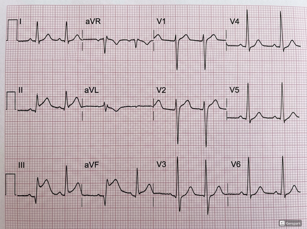
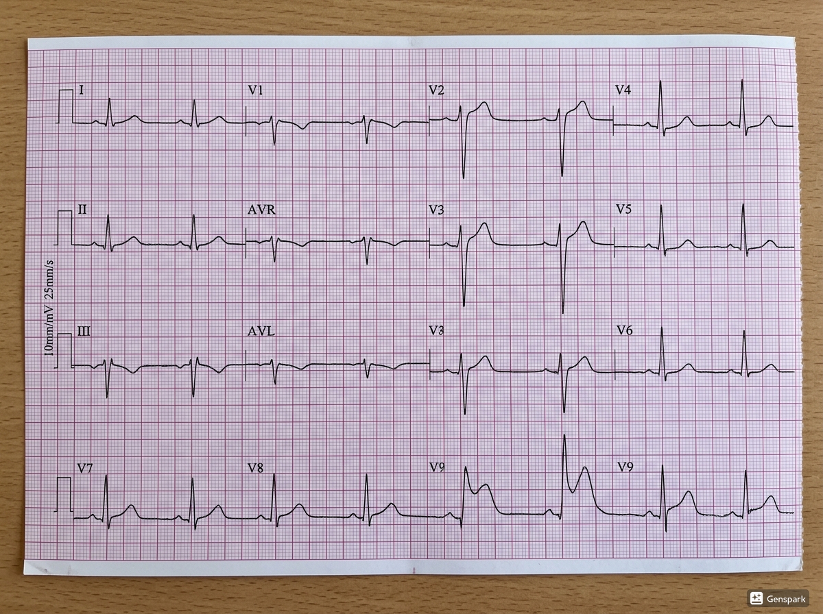
Posterior MI is suggested by a characteristic pattern in leads V1 to V3: horizontal ST depression, tall broad R waves, upright T waves, and often a dominant R wave in V2 with an R/S ratio greater than 1. These findings are the reciprocal equivalents of the usual STEMI features. In effect, posterior ST elevation becomes anterior ST depression, posterior Q waves become anterior R waves, and terminal T-wave inversion becomes an upright T wave.
For learners, the easiest phrase to remember is this: posterior STEMI is the mirror image of an anterior tracing. When the ST depression in V1 to V3 is accompanied by unexpectedly tall R waves and upright T waves, the tracing should make you think of posterior infarction rather than routine subendocardial ischaemia.
Why ST depression in V1-V3 can mean STEMI
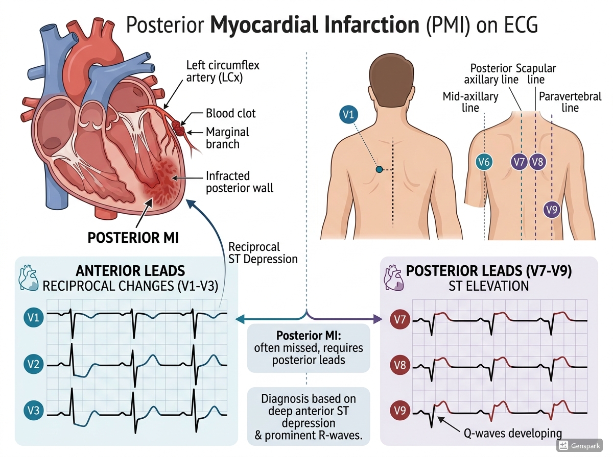
The explanation is anatomical and electrical. The anteroseptal leads are recording posterior myocardial activity from the opposite side of the heart. As a result, the usual posterior STEMI injury pattern is inverted when seen from the front. ST elevation becomes ST depression, Q waves become R waves, and T-wave inversion becomes an upright T wave. Once you understand that mirror relationship, the pattern becomes far easier to recognize and much harder to forget.
This is why isolated anterior ST depression should never be dismissed automatically. In the right clinical context, especially when the morphology is horizontal and accompanied by tall R waves, it should raise immediate concern for posterior MI.
How to confirm posterior MI with V7-V9
Posterior MI is confirmed by recording posterior leads V7, V8, and V9. These leads are placed across the posterior chest wall in the same horizontal plane as V6: V7 at the posterior axillary line, V8 at the tip of the scapula, and V9 at the paraspinal region. ST elevation in these leads confirms posterior infarction. Teaching sources emphasize that even 0.5 mm of ST elevation in V7 to V9 is sufficient for diagnosis.
This is a high-yield bedside step. When you see suspicious ST depression in V1 to V3 with posterior features, record posterior leads rather than debating endlessly whether the pattern “looks ischemic enough.” Posterior leads turn a hidden infarct into a visible one.
Posterior MI rarely comes alone
Posterior myocardial infarction accompanies 15% to 20% of STEMIs and usually occurs with inferior or lateral infarction. Isolated posterior MI is less common. This is another practical clue: if a patient has inferior or lateral STEMI plus suspicious anterior ST depression in V1 to V3, think about posterior extension. That often means a larger infarct territory and a higher-risk situation than a simple isolated territory pattern.
Because posterior MI often travels with other infarct territories, the whole ECG must be read as a map. Do not focus only on V1 to V3. Look for inferior ST elevation, lateral involvement, or other clues that the infarct extends beyond one wall.
Posterior MI vs anterior ischemia: how not to get fooled
Not every ST depression pattern in V1 to V3 is posterior MI. Anterior subendocardial ischaemia, diffuse demand ischaemia, bundle branch block, or technical issues may also alter the tracing. What makes posterior MI different is the specific combination of horizontal ST depression, tall broad R waves, dominant R-wave progression, and upright T waves in the early precordial leads. Those tall R waves are especially important because they act as the Q-wave equivalent on the opposite side of the heart.
In practical teaching terms: if the ST depression is accompanied by a surprisingly positive, broad, dominant R wave in V2 or V3, posterior infarction should move high up your differential.
Why timing and repeat ECGs still matter
In suspected acute coronary syndrome, the ECG should be obtained and interpreted within 10 minutes of presentation. An initial nondiagnostic ECG does not exclude ACS because the changes can be dynamic. This is particularly important in posterior MI, where the pattern may be subtle or evolving and where specialized leads may be needed to reveal the diagnosis.
Guidance emphasizes comparing with prior ECGs, repeating the tracing during the emergency department course, and obtaining posterior leads when suspicion remains. If recurrent chest pain or clinical deterioration occurs, the threshold for repeat ECGs should be low.
A practical bedside checklist
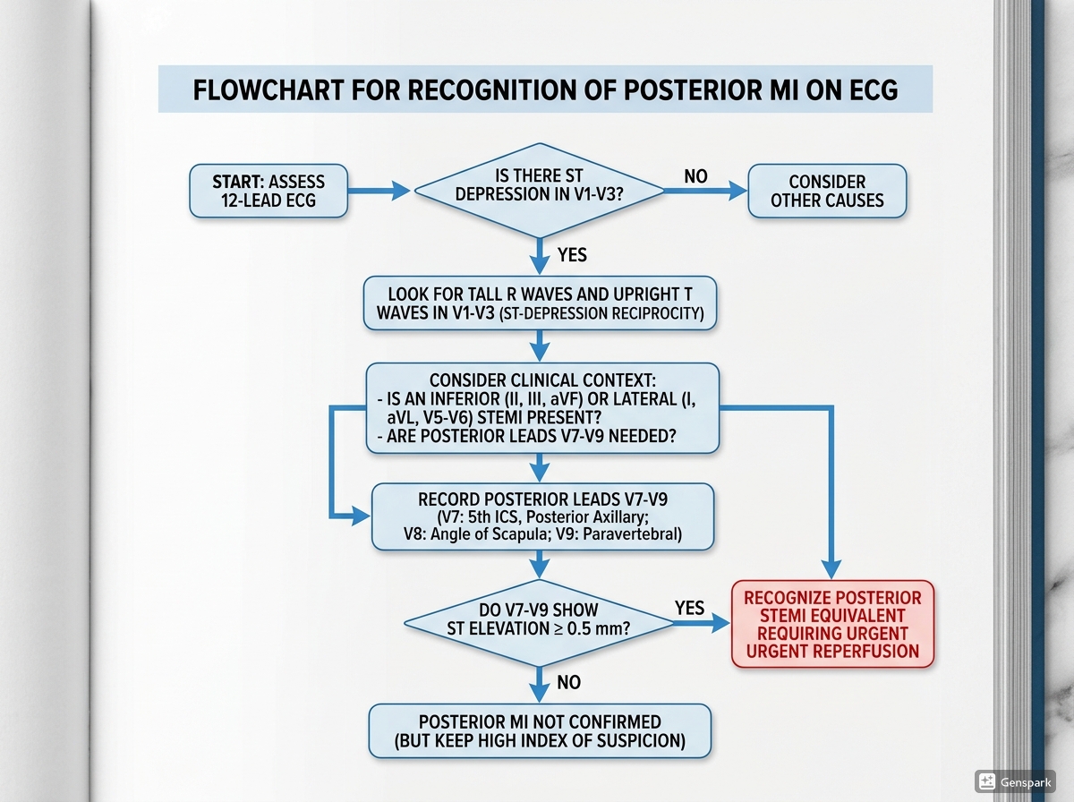
When you suspect posterior MI, use this quick sequence:
- Look for horizontal ST depression in V1 to V3.
- Check for tall, broad R waves and upright T waves in the same leads.
- Ask whether the pattern could be a mirror image of posterior STEMI rather than ordinary anterior ischaemia.
- Inspect the inferior and lateral leads for associated STEMI patterns.
- Record posterior leads V7 to V9 when the pattern is suspicious.
- Escalate urgently because posterior MI is a STEMI-equivalent pattern that may require reperfusion.
Common mistakes to avoid
- Assuming ST depression always means NSTEMI or diffuse subendocardial ischaemia.
- Ignoring tall R waves in V1 to V3 when interpreting anterior ST depression.
- Failing to obtain posterior leads V7-V9 in a suspicious case.
- Missing associated inferior or lateral infarction that makes posterior extension more likely.
- Stopping after one nondiagnostic ECG even though symptoms continue or worsen.
Frequently asked questions
What is the classic ECG pattern of posterior MI?
Horizontal ST depression in V1-V3 with tall broad R waves, upright T waves, and often a dominant R wave in V2.
Why can ST depression in V1-V3 represent STEMI?
Because posterior wall injury is viewed from the opposite side, so the usual STEMI pattern appears as a mirror image in the anterior leads.
How do you confirm posterior MI?
Record posterior leads V7-V9. ST elevation in these leads confirms the diagnosis.
Does posterior MI usually occur alone?
Not usually. It often accompanies inferior or lateral infarction, while isolated posterior MI is less common.
Why is posterior MI important not to miss?
Because it may represent an occlusive infarction needing urgent reperfusion, yet it can be mistaken for NSTEMI if the mirror-image pattern is not recognized.
Key takeaways
- Posterior MI is often hidden on the standard 12-lead ECG and may present as anterior ST depression rather than obvious ST elevation.
- Key clues in V1-V3 are horizontal ST depression, tall broad R waves, upright T waves, and dominant R-wave progression.
- Posterior MI is confirmed by ST elevation in posterior leads V7-V9.
- Posterior infarction often occurs with inferior or lateral STEMI rather than in isolation.
- Suspicious anterior ST depression with tall R waves should trigger posterior lead recording and urgent reassessment.
- Posterior MI should be treated as a STEMI-equivalent pattern when clinically appropriate.
References
- Posterior Myocardial Infarction – ECG Library Diagnosis.
- The ST Segment – ECG Library Basics.
- 2025 ACC/AHA/ACEP/NAEMSP/SCAI Guideline for the Diagnosis and Risk Classification of Acute Coronary Syndromes.
- Thygesen K, Alpert JS, Jaffe AS, et al. Fourth Universal Definition of Myocardial Infarction.