Cardiac Axis Deviation on ECG Explained
Description: Learn how to assess cardiac axis on ECG using leads I and aVF, understand normal axis, left axis deviation, right axis deviation, and extreme axis deviation, and know the common clinical causes.

Quick Answer
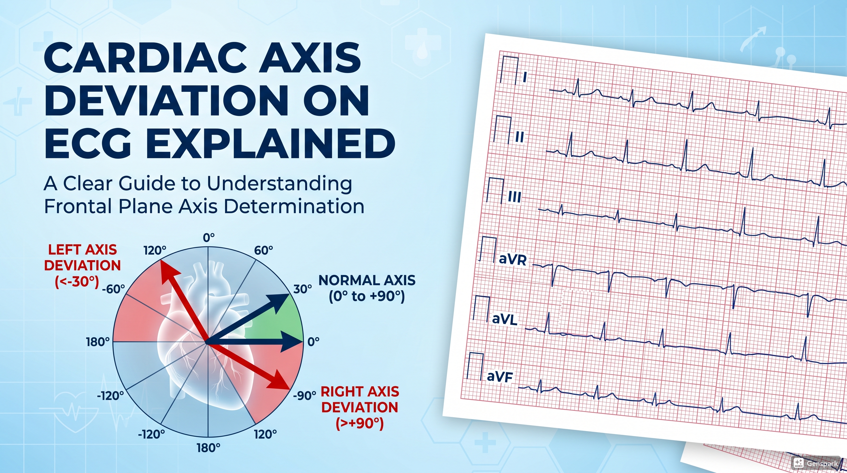
Cardiac axis on ECG refers to the overall direction of ventricular depolarization in the frontal plane. In practical bedside teaching, normal QRS axis is usually about -30° to +90°. Left axis deviation is less than -30°, right axis deviation is greater than +90°, and extreme axis deviation lies between -90° and 180°. The quickest way to estimate axis is to look at the QRS polarity in leads I and aVF, then place the axis into the correct quadrant.
Introduction
Many medical students learn rate, rhythm, and intervals early in ECG interpretation, but axis is often left until later or treated as something abstract. In reality, cardiac axis is one of the most useful parts of a structured ECG read. It can help you recognize fascicular block, chamber hypertrophy, ventricular rhythms, lead misplacement, and important cardiopulmonary disease patterns.
The reason axis feels confusing at first is that it is usually taught as geometry before it is taught as a practical bedside skill. Most junior learners do not actually need advanced mathematics to interpret axis. What they need is a simple framework that can be applied quickly and safely on real ECGs.
This article explains cardiac axis deviation step by step in a mixed educational style suitable for broader traffic, while still being clinically useful for medical students and junior doctors.
What Is Cardiac Axis on ECG?
The electrical axis of the heart reflects the average direction of ventricular depolarization during ventricular contraction. In simpler terms, it tells you which way the main ventricular electrical impulse is pointing in the frontal plane. Because the left ventricle usually dominates depolarization, the normal axis generally points leftward and downward.
Axis is assessed using the limb leads rather than the chest leads. This is important because the limb leads view the heart in the frontal plane, which is exactly the plane in which axis is defined.
Why Axis Matters Clinically
Axis is not just an academic number. It can support the diagnosis of left anterior fascicular block, left posterior fascicular block, ventricular hypertrophy, inferior infarction, right ventricular strain, pulmonary disease, ventricular rhythms, and even technical problems such as limb lead misplacement.
If you consistently ignore axis, you may still recognize some obvious ECGs, but you will miss useful clues that make your interpretation sharper and more complete.
Normal Axis Range
In everyday ECG teaching, normal QRS axis is usually described as approximately -30° to +90°. Some sources simplify the normal range slightly differently, but this is the standard practical range used in many bedside and exam-focused resources.
Abnormal axis ranges
- Normal axis: about -30° to +90°
- Left axis deviation: less than -30°
- Right axis deviation: greater than +90°
- Extreme axis deviation: between -90° and 180°

The Easiest Way to Estimate Axis: Leads I and aVF
The fastest practical method is the quadrant method using lead I and lead aVF. You do not need to calculate the exact degree every time. First, inspect whether the QRS complex in each lead is predominantly positive or predominantly negative.
How to use the quadrant method
- Lead I positive and aVF positive = normal axis
- Lead I positive and aVF negative = possible left axis deviation
- Lead I negative and aVF positive = right axis deviation
- Lead I negative and aVF negative = extreme axis deviation
This method is fast, reliable, and ideal for beginners. Once you are comfortable with it, you can refine the interpretation further using lead II or the full hexaxial reference system if needed.

A Useful Refinement: What About Lead II?
If lead I is positive and aVF is negative, the axis may be either normal but leftward or truly left axis deviation. This is where lead II becomes helpful. If lead II is still positive, the axis is usually still within the normal range. If lead II is negative, true left axis deviation is more likely.
This small extra step helps avoid overcalling left axis deviation in borderline cases.
What Does a Positive or Negative QRS Mean?
A lead is considered positive if the overall QRS deflection is mostly above the baseline. It is considered negative if the overall QRS deflection is mostly below the baseline. In practice, you are judging the net direction of the QRS, not focusing on every tiny deflection.
If the QRS is nearly equal above and below the baseline, it may be considered equiphasic or isoelectric. That can be useful for more detailed axis estimation, but beginners should first master the simple quadrant method.
Left Axis Deviation on ECG
Left axis deviation means the QRS axis is more negative than -30°. In the simple bedside method, this usually appears as a predominantly positive QRS in lead I and a predominantly negative QRS in aVF, often with a negative QRS in lead II as well.
Common causes of left axis deviation
- Left anterior fascicular block
- Left ventricular hypertrophy
- Left bundle branch block
- Inferior myocardial infarction
- Ventricular pacing or ventricular ectopy
- Pre-excitation in some cases
Among these, left anterior fascicular block is one of the classic high-yield causes that students and junior doctors should know well.

Right Axis Deviation on ECG
Right axis deviation means the QRS axis is greater than +90°. In the simple quadrant method, this usually appears as a predominantly negative QRS in lead I and a predominantly positive QRS in aVF.
Common causes of right axis deviation
- Right ventricular hypertrophy
- Acute right ventricular strain, such as pulmonary embolism
- Chronic lung disease, including COPD
- Pulmonary hypertension
- Left posterior fascicular block
- Dextrocardia or limb lead problems in some cases
In younger patients, a relatively rightward axis may sometimes be normal, and in paediatric ECGs rightward axis is especially common. Clinical context matters.
Extreme Axis Deviation
Extreme axis deviation, sometimes called the northwest axis, is present when both lead I and aVF are predominantly negative. This pattern is uncommon. When you see it, think carefully about technical issues such as limb lead misplacement, or serious pathological causes such as ventricular tachycardia if the rhythm is wide-complex and clinically concerning.
How the Hexaxial Reference System Helps
The hexaxial reference system is the full frontal-plane map of the limb leads. It allows you to estimate axis more precisely by using the angles assigned to leads I, II, III, aVR, aVL, and aVF. Although this is useful, beginners do not need to calculate exact angles on every ECG. In routine practice, the quadrant method gives a fast and clinically effective answer in most cases.
The hexaxial system becomes more useful when you want to refine borderline cases or understand why particular leads behave the way they do.
Normal Axis vs Axis Deviation on a Real ECG
When you look at an ECG with normal axis, the overall QRS is usually positive in lead I and positive in aVF. In contrast, a tracing with left axis deviation will usually keep a positive QRS in lead I but show a negative QRS in aVF. Recognizing this pattern repeatedly in real tracings is one of the best ways to build confidence.

Common Beginner Mistakes
One common mistake is trying to find the exact numerical axis before first deciding whether the axis is broadly normal, leftward, rightward, or extreme. Start simple. Another common mistake is judging a single large spike rather than the net QRS direction.
A second frequent problem is forgetting that limb lead misplacement can create bizarre axis patterns. If the ECG looks inconsistent with the clinical picture, think about technical error before diagnosing rare pathology.
Students also sometimes overinterpret axis in isolation. Axis is one clue, not the whole diagnosis. A leftward axis alone does not automatically mean left anterior fascicular block unless the rest of the ECG pattern supports it.
A Practical Memory Aid
If you want a quick mental shortcut, remember this:
- Lead I positive, aVF positive = normal
- Lead I positive, aVF negative = leftward
- Lead I negative, aVF positive = rightward
- Lead I negative, aVF negative = extreme
Then use lead II when needed to decide whether the leftward pattern is truly left axis deviation or still within the normal border zone.
FAQ
What is the normal cardiac axis on ECG?
In practical adult ECG interpretation, the normal QRS axis is usually about -30° to +90°.
How do you identify left axis deviation quickly?
Look for a positive QRS in lead I and a negative QRS in aVF. Then check lead II if needed to confirm true left axis deviation.
What causes right axis deviation?
Common causes include right ventricular hypertrophy, pulmonary hypertension, pulmonary embolism, chronic lung disease, and left posterior fascicular block.
What does extreme axis deviation suggest?
Extreme axis deviation may suggest limb lead misplacement, ventricular rhythms, or other significant pathology depending on the ECG pattern and clinical context.
Do chest leads determine cardiac axis?
No. Cardiac axis is assessed primarily using the limb leads because axis is defined in the frontal plane.
Key Takeaways
Cardiac axis on ECG is the average direction of ventricular depolarization in the frontal plane. For most practical purposes, normal axis is about -30° to +90°, left axis deviation is less than -30°, right axis deviation is greater than +90°, and extreme axis deviation lies between -90° and 180°.
For medical students and junior doctors, the best first method is the quadrant method using leads I and aVF. It is fast, simple, and clinically useful. Once you master that, axis becomes much less intimidating and much more valuable in everyday ECG interpretation.
References
- American Heart Association. Electrocardiogram (EKG or ECG).
- LITFL. ECG Axis Interpretation.
- ECG Waves. The Electrical Axis of the Heart (Heart Axis).
- ECG Waves. Reference Values for Adult ECG.