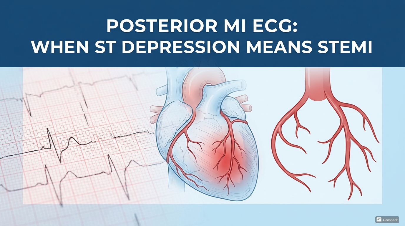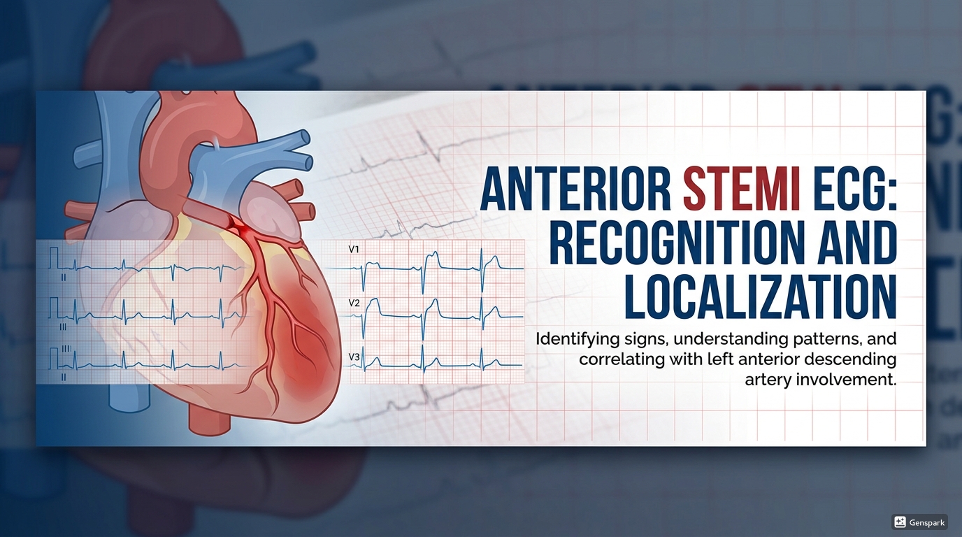
Anterior STEMI ECG: Recognition and Localization
Description: Learn how to recognize anterior STEMI on ECG, localize septal, anterior, anteroseptal, and anterolateral patterns, and spot high-risk clues suggesting proximal LAD occlusion.
|
Quick answer: Anterior STEMI is classically recognized by ST-segment elevation in the precordial leads, especially V1 to V4 or V2 to V5 depending on the exact territory involved, often with reciprocal ST depression in the inferior leads. Localization is based on which precordial and lateral leads are involved: septal infarction affects V1-V2, anterior infarction V2-V5, anteroseptal V1-V4, anterolateral V3-V6 with I and aVL, and extensive anterior infarction V1-V6 with I and aVL. These patterns usually reflect LAD occlusion and may hint at how proximal the lesion is. |
Anterior STEMI is one of the most dangerous and high-yield ECG diagnoses in acute cardiology. It often reflects occlusion of the left anterior descending artery, threatens a large myocardial territory, and carries a worse prognosis than many other infarct locations because of the amount of left ventricular muscle at risk. For medical students and junior doctors, this is one of the ECG patterns you must be able to recognize quickly and describe clearly.
The challenge is that “anterior STEMI” is not a single fixed picture. The exact lead pattern can help localize the infarct as septal, anterior, anteroseptal, anterolateral, or extensive anterior. These distinctions are useful because they can hint at the infarct size, the likely LAD segment involved, and the urgency of anticipating complications such as cardiogenic shock, arrhythmia, or extensive left ventricular dysfunction. The goal is not to memorize a list mechanically, but to understand how the precordial and high lateral leads map onto the anterior wall and septum.
What is anterior STEMI?
Anterior STEMI refers to acute transmural infarction involving the anterior wall of the left ventricle, usually because of acute LAD occlusion. On the ECG, it appears as ST-segment elevation in the precordial leads. These leads look directly at the anterior and septal surfaces of the heart, which is why acute injury in this region produces such striking precordial changes.
Because the LAD supplies a large territory, anterior STEMI often represents a large infarct burden. Compared with smaller infarct territories, this pattern is more strongly associated with pump failure, shock, and poor outcomes. That is why early recognition matters so much.
The core ECG features of anterior STEMI
The classic ECG features are ST-segment elevation in the precordial leads with later Q-wave formation. Hyperacute T waves may appear before the ST elevation becomes obvious. Reciprocal ST depression often appears in the inferior leads, especially III and aVF. If the infarction is large or more extensive, high lateral leads such as I and aVL may also be involved.
For beginners, a safe first-pass question is: are the precordial leads showing convincing contiguous ST elevation? If yes, the next question becomes localization. Which leads are most involved, and do high lateral leads participate? That is where the naming system becomes useful.

How to localize anterior STEMI by lead distribution
A practical way to localize anterior STEMI is to name the infarct pattern according to the leads showing maximal ST elevation. Septal infarction involves V1 and V2. Anterior infarction more broadly involves V2 to V5. Anteroseptal infarction involves V1 to V4. Anterolateral infarction involves V3 to V6 together with leads I and aVL. Extensive anterior or extensive anterolateral infarction may involve V1 to V6 plus I and aVL.
These patterns are not just labels for exams. They help you communicate clearly and think about infarct size. As a rule, the more widespread the ST elevation across the precordial and lateral leads, the larger and more proximal the affected territory is likely to be.

Septal, anterior, anteroseptal, and anterolateral patterns
Septal STEMI
Septal STEMI shows maximal ST elevation in V1 and V2. Because these leads reflect the interventricular septum, this pattern suggests septal involvement and can point toward a lesion affecting septal branches of the LAD.
Anterior STEMI
Anterior STEMI more broadly affects V2 to V5 and reflects infarction in the anterior wall. This is the pattern many learners think of first when they hear “LAD occlusion.”
Anteroseptal STEMI
Anteroseptal STEMI involves V1 to V4 and combines septal and anterior territory. This usually suggests a lesion affecting the LAD territory more proximally than a purely limited anterior or septal pattern.

Anterolateral and extensive anterior STEMI
When ST elevation extends into V3 to V6 and the high lateral leads I and aVL, the pattern becomes anterolateral. If V1 to V6 plus I and aVL are involved, this suggests extensive anterior or extensive anterolateral infarction. These widespread patterns imply a larger territory at risk and should immediately raise concern for a large proximal LAD lesion and worse haemodynamic consequences.
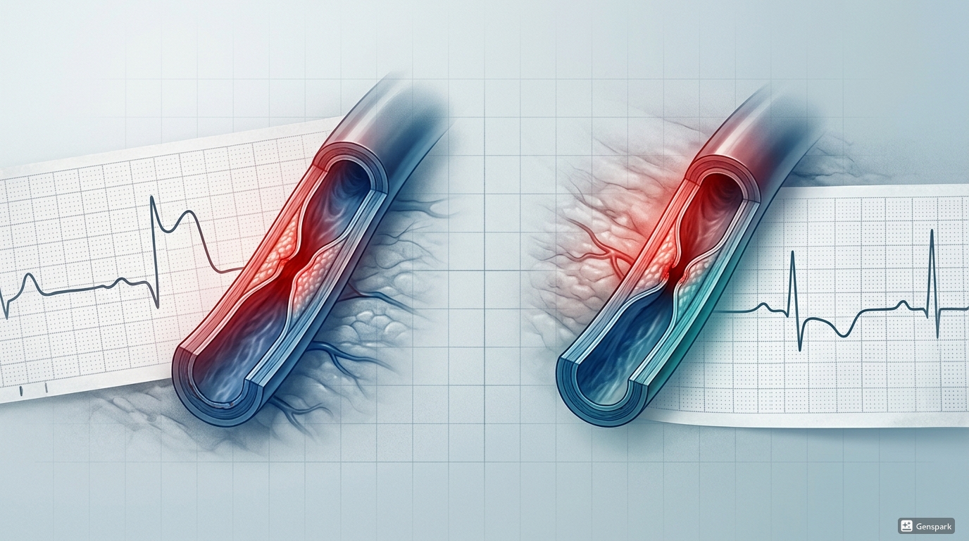
Clues to proximal LAD occlusion
Some ECG findings suggest that the LAD occlusion lies proximal to important branches such as the first septal branch or first diagonal branch. Teaching sources emphasize that ST elevation in aVR, marked ST elevation in V1, complete right bundle branch block, or involvement of the high lateral leads can point toward a more proximal lesion. When the precordial ST elevation is widespread and the lateral leads also participate, think about a larger anterior infarction rather than a small focal process.
These are clues, not perfect angiographic rules. Still, for a junior doctor, recognizing the concept is valuable: the more proximal the LAD lesion, the more extensive the territory at risk may be.
Reciprocal changes and why they matter
Anterior STEMI often produces reciprocal ST depression in the inferior leads, especially III and aVF. These reciprocal changes help support the diagnosis, particularly when the precordial elevation is not dramatic. As in other STEMI patterns, reciprocal changes are useful because they strengthen the case that the tracing reflects acute infarction rather than a benign mimic.
A good bedside habit is to examine the inferior leads whenever you suspect anterior STEMI. Inferior reciprocal depression is not mandatory, but when present it adds confidence and can make subtle anterior STEMI easier to recognize.
High-risk anterior ischemic patterns to know
Not all dangerous LAD-related patterns present as classic anterior ST elevation. High-risk anterior ischaemic patterns include Wellens syndrome, De Winter T waves, and widespread depression with ST elevation in aVR suggesting left main or severe proximal disease. These patterns remind clinicians that the absence of classic precordial STEMI criteria does not always mean the patient is low risk. However, the present article focuses on classic anterior STEMI localization rather than those STEMI equivalents.
Why timing and repeat ECGs still matter
In suspected ACS, the ECG should be obtained and interpreted within 10 minutes of presentation. This is especially important in anterior STEMI because early reperfusion decisions are time-sensitive and the territory at risk is often large. At the same time, an initially nondiagnostic ECG does not rule out ACS. ECG abnormalities are dynamic, and repeat ECGs should be obtained when symptoms persist, recur, or the clinical condition changes.
This matters clinically because some patients who ultimately have STEMI do not meet diagnostic ECG criteria on the first tracing. If the symptoms fit and the suspicion remains high, repeat the ECG and compare with previous tracings whenever possible.

A practical bedside checklist
When you suspect anterior STEMI, work through the ECG in this order:
- Look for contiguous ST elevation in the precordial leads.
- Identify which leads are most involved: V1-V2, V1-V4, V2-V5, or V3-V6 with I and aVL.
- Check for inferior reciprocal ST depression in III and aVF.
- Decide whether the pattern is septal, anterior, anteroseptal, anterolateral, or extensive anterior.
- Look for clues suggesting a proximal LAD lesion, such as widespread involvement or high lateral extension.
- Escalate quickly because anterior STEMI usually represents a large territory at risk.
Common mistakes to avoid
- Calling all precordial ST elevation “anterior STEMI” without localizing the involved territory.
- Missing subtle reciprocal inferior changes that support the diagnosis.
- Underestimating the prognostic importance of widespread precordial and lateral lead involvement.
- Failing to repeat the ECG when symptoms are typical but the first tracing is nondiagnostic.
- Forgetting that some LAD occlusions present with STEMI equivalents rather than classic ST elevation.
Frequently asked questions
Which leads define classic anterior STEMI?
Anterior STEMI usually shows ST elevation in the precordial leads, especially V2 to V5, although the exact pattern depends on whether the infarct is septal, anterior, anteroseptal, or anterolateral.
What is the difference between septal and anteroseptal STEMI?
Septal STEMI is centered in V1-V2, while anteroseptal STEMI usually involves V1-V4, reflecting broader LAD territory involvement.
Why is anterior STEMI considered high risk?
Because it often reflects LAD occlusion and threatens a large amount of left ventricular myocardium, increasing the risk of heart failure, shock, and large infarct size.
What reciprocal changes are common in anterior STEMI?
Reciprocal ST depression is often seen in inferior leads, especially III and aVF.
Can the first ECG be nondiagnostic in anterior STEMI?
Yes. ECG abnormalities can evolve, so repeat ECGs are important if symptoms persist or suspicion remains high.
Key takeaways
- Anterior STEMI usually reflects LAD occlusion and often threatens a large myocardial territory.
- Core ECG findings are precordial ST elevation, often with later Q waves and inferior reciprocal depression.
- Lead patterns help localize the infarct as septal, anterior, anteroseptal, anterolateral, or extensive anterior.
- Widespread precordial and high lateral involvement suggests a larger, potentially more proximal LAD lesion.
- Anterior STEMI is high risk and should be recognized rapidly as a reperfusion emergency.
- A nondiagnostic first ECG does not exclude STEMI if symptoms remain concerning, so repeat ECGs matter.
References
- Anterior Myocardial Infarction – ECG Library Diagnosis.
- MI Localization – ECG Library Basics.
- 2025 ACC/AHA/ACEP/NAEMSP/SCAI Guideline for the Diagnosis and Risk Classification of Acute Coronary Syndromes.
- Thygesen K, Alpert JS, Jaffe AS, et al. Fourth Universal Definition of Myocardial Infarction.