A physical cardiac examination is essential for the assessment of heart structure and function and the detection of any abnormalities. Physical cardiac examination is not restricted to auscultation of heart sounds using a stethoscope, as palpation of arterial pulse, measurement of blood pressure, inspection of general appearance, and examination of the neck, extremities, mouth, skin, and nails have a critical role.
Neck examination through assessing the jugular vein pulse and identifying any distention can aid in the evaluation of central vein pressure and the diagnosis of valvular heart diseases and atrial arrhythmia.
Jugular vein pressure (JVP)
Jugular vein pressure is assessed using the right internal jugular vein, which is located at the right side of the nick underneath the sternocleidomastoid muscle between the ear, the lobe, and the medial clavicle. The patient must be set at 30°–45° elevated from the supine position while slightly turning the head to the left.
When the jugular vein is assessed, two pulses will be visualized for each heartbeat, but they are not palpable pulsations. Also, the pressure will be temporarily elevated as a hepatojugular reflex in the case of pressing on the right upper quadrant of the abdomen. In addition, the jugular vein will normally collapse during inspiration.
Jugular vein pressure height is measured vertically from the sternal angle to the intersection point with the horizontal level of jugular vein pulsation, which is shorter than 3cm under normal conditions. Then, five is added to the jugular vein height to convert it to jugular vein pressure in cm H2O units.
JPV and atrial waveforms
Jugular vein pressure can be used as an indirect measurement of right atrium pressure since non-oxygenated blood moves directly through it to the superior vena cava and right atrium. As a result, several waves are formed and reflect atrial conditions.
- A wave: represents atrial contraction
Atrial contraction increases atrial pressure, which leads to blood movement through the tricuspid valve into the right ventricle, as well as blood movement back into the jugular vein, which increases its pressure.
- C wave: represents ventricular contraction
Ventricular systole results in elevated pressure and bulging of the closed tricuspid valve, which leads to back pressure at the right atrium and jugular vein.
- X descent: represents atrial relaxation
Due to atrial relaxation, atrial pressure is dropped, which indirectly causes a drop in jugular vein pressure.
- V wave: represents venous atrial filling
Atrium venous filling increases the atrial pressure and leads to blood backflow into a jugular vein as the tricuspid valve is closed at this phase.
- Y descent: represents ventricular filling
In this phase, the tricuspid valve is opened and ventricular filling is accelerated, so atrium and jugular vein pressure are falling.
Cardiac conditions related to JVP and atrial waves
- Increased jugular vein pressure during inspiration (Kussmaul's sign) is associated with an abnormality in atrial filling due to constrictive pericarditis or right ventricular hypertrophy.
- Decreased Jugular vein pressure is associated with hypovolemia.
- A wave absence and X descent attenuation are observed in atrial fibrillation cases when atrial contractions are not coordinated and the atrium becomes unable to relax.
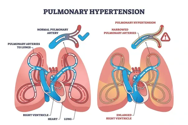
- Large A wave and attenuated Y descent are observed when blood is not moved smoothly from the atrium to the ventricle because of right ventricular hypertrophy, pulmonary hypertension, or tricuspid valve stenosis.
- Cannon A wave is observed when the atrial and ventricles contract simultaneously due to third-degree atrioventricular heart block, which impedes blood movement from the atrium to the ventricle.
- A large V wave is observed with tricuspid valve regurgitation, which is associated with X descent absence and CV wave formation due to increased blood volume in the atrium.
- Y descent attenuation is observed due to the inability of the atrium to relax while ventricles are in diastole, which occurs due to elevated pressure on the right atrium as in the Cardiac Tamponade.
- Prominent Y descent (Friedreich's sign) is observed if the ventricle is unable to expand, as in constrictive pericarditis.
References


.webp)
.webp)
.webp)
.webp)
.webp)

.webp)