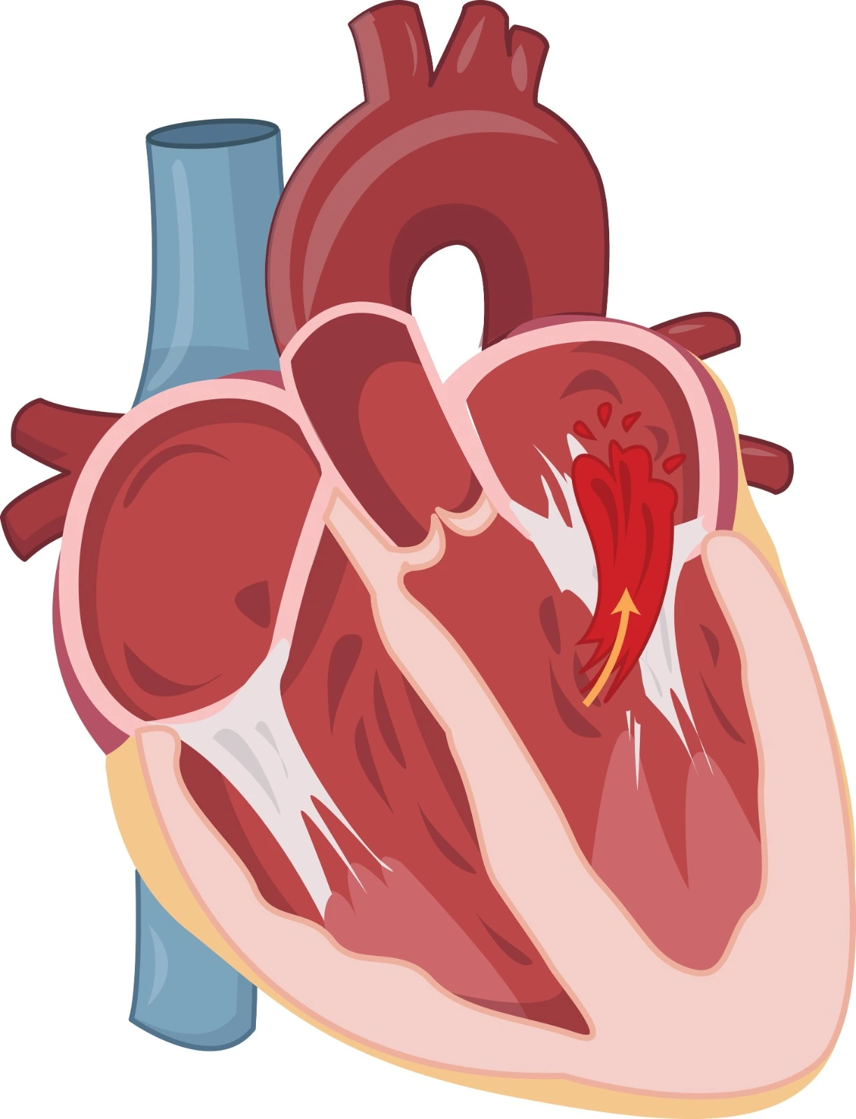
A cardiac electrical signal normally originates from the right atrium Sinoatrial (SA) node and is associated with atrial contraction, which is represented by a P wave at the ECG, is then transmitted to the atrioventricular (AV) node between the right atrium and ventricle, is then conducted to bundles of his and moves along the ventricular wall toward the heart apex and through Purkinje fibers, leading to ventricle contraction, which is represented by the QRS complex on the ECG.
Supraventricular tachycardia (SVT) is a common type of cardiac arrhythmia that affects normal electrical impulse generation and conduction. The nomenclature is related to the fact that this type of arrhythmia is initiated in the atrial region. It is associated with an elevated heart rate, and the patient may be asymptomatic or complain of palpitation, shortness of breath, chest pain, diaphoresis, fatigue, and fainting or syncope.
Differential diagnosis
where the heart rate is elevated and the heart rhythm is in a regular sinus rhythm. It is usually physiological and related to secondary conditions such as sepsis, hypovolemia, heart failure, anemia, hyperthyroidism, and medications with beta-receptor stimulation effects.
It is diagnosed if the heart rate is elevated, the heart rhythm is irregularly irregular, the QRS interval is narrowed to <120 ms and the P wave is absent.
It is diagnosed when the heart rate is markedly elevated, the heart rhythm is usually regular, the QRS interval is narrowed to <120 ms and a saw-tooth pattern is observed.
It is usually identified through ECG; when the heart rate is elevated, heart rhythm is regular, and the QRS interval is narrowed to <120 ms. In some cases, the QRS interval may be wide if accompanied by heart block or accessory pathway conduction.
Etiology and Pathophysiology
In supraventricular tachycardia, electrical signals may be fired from an abnormal origin, conducted through abnormal pathways, or retrogradely conducted back to the atrium instead of the normal unidirectional anterograde electrical pathway.
-
Atrioventricular Nodal Reentrant Tachycardia (AVNRT)
It is one of the most common causes among adults and females. AV nodal tissues usually have one conduction pathway, where the signal moves in a unidirectional way down to the bundle of his. Arrhythmia related to the reentry mechanism is dependent on the reentry circuit, which consists of two electrically different pathways along with premature atrial signals.
At typical AVNRT, a first fast-conduction pathway with a long refractory period gets activated at its distal part by a premature signal conducted through a second slow-conduction and short-refractory pathway, which also activates the ventricle, then the signal will move up to the atrium through the first pathway, then a new premature atrial signal will activate the second pathway, and the cycle will be repeated. This resulted in the simultaneous contraction of the atria and ventricles and was represented by a longer PR interval and P wave after the QRS complex.
Atypical AVNRT may occur when an electrical signal is conducted from the first fast-conduction pathway and then from the second slow-conduction pathway up to the atrium. IZt is represented by a longer RP compared to PR.
-
Atrioventricular Reciprocating Tachycardia (AVRT)
It is common at young ages and among males. In this case, the accessory pathway is connected to the normal atrioventricular pathway to form a reentry circuit. It is associated with 100–250 beats per minute.
Orthodromic AVRT is a common type, where extra systolic impulses move down the normal AV pathway and then in a retrograde manner up to the atrium through an accessory pathway with a long refractory period. It is associated with Wolf Parkinson White Syndrome (WPWS) and other concealed accessory pathways. It is represented by a narrow QRS, a short RP, and an inverted P wave after the QRS.
Antidromic AVRT is a very rare type where the electrical signal is conducted in an anterograde manner through an accessory pathway and then to a normal AV pathway up to the atrium and down for ventricle pre-excitation, which is associated with Wolf Parkinson White Syndrome (WPWS). It is reflected as a wide QRS, a long RP, and an inverted P wave after the QRS.
■ Wolf Parkinson White Syndrome (WPWS) is usually represented as a delta wave (upstroke to QRS complex) on the ECG. If electrical signal firing becomes faster through accessory pathways, it may participate in atrial fibrillation and flutter and increase ventricular fibrillation and sudden death risk.
In this case, tachyarrhythmia is related to increased tissue automaticity in the atrium region, resulting in ectopic firing and an elevated heart rate that ranges from 100 to 250 bpm. Common ectopic origins include the left or right atrium, superior vena cava, non-coronary aortic cusp, and rarely a hepatic vein. It may have occurred in people with normal or abnormal heart conditions. Also, electrolyte and catecholamine disturbances and some medications are considered precipitating factors.
References


.webp)
.webp)
.webp)
.webp)
.webp)
.webp)

.webp)