The aortic valve is a semilunar valve consisting of three collagen leaflets or cusps: right coronary, left coronary, and non-coronary, which are connected to the aortic root by a fibrous virtual ring called the annulus. It is located between the left ventricle and the aorta. It is opened during heart systole to allow oxygenated blood ejection through the aorta to the whole body, and it is closed during heart diastole to allow left ventricle oxygenated blood filling without any inward or backward leakage.
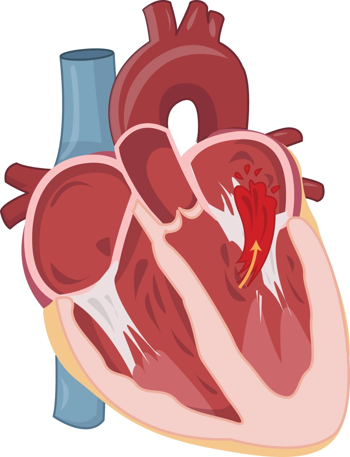
Aortic valve Regurgitation is one of the valvular heart disorders that affects its normal structure and function; it occurs when the aortic valve abnormally allows the oxygenated blood to leak backward from the aorta to the left ventricle during cardiac diastole.
Underlying Causes
Aortic regurgitation prevalence is higher in males and increases with aging. Disorders that affect any valve structure acutely or chronically could cause aortic regurgitation.
■ Active infectious endocarditis (IE), as seen in large vegetation, may lead to a perivalvular abscess that affects valve leaflets through destruction or perforation.
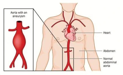
■ Acute Aortic dissection in the proximal ascending part may affect valve root leaflet suspension and could lead to prolapse.
■ Acute trauma may occur due to cardiac procedures, such as catheterization or device insertion, and lead to valve cusp rupture.
■ Prosthetic valve dysfunction is caused by endocarditis or structural destruction due to valve fibrosis or calcification. When the valve and annulus are not fully sealed together, it could lead to a valvular rupture.
■ Degenerative aortic regurgitation, where abnormality of aortic valve structure is due to defect in collagen and elastic fiber formation and organization.
■ Bicuspid Aortic valve is a common congenital disorder where aortic valve cusps are only two leaflets; they are usually asymmetrical due to the fusion of two cusps together, and it affects the aorta and leads to chronic aortic regurgitation.
■ Rheumatic fever and rheumatoid heart disease may develop due to one or more episodes of rheumatic fever. In this case, an immunological response to Streptococcus pyogenes proteins may lead to an autoimmune attack on cardiac proteins, which leads to inflammation and fibrosis of the aortic valve leaflet and chronic regurgitation.
■ Ankylosing spondylitis is a rheumatic disease where chronic inflammation can affect the whole aortic root.
■ Rheumatoid arthritis is an inflammatory disease.
■ Systemic lupus erythematosus (SLE) is an autoimmune disease.
■ Marfan syndrome and Ehlers-Danlos syndrome are genetic disorders that affect connective tissues and induce dilation in the aortic valve root, aneurysm, or dissection in the aorta.
Symptoms and Cardiac Complications
Aortic regurgitation symptoms are dependent on the underlying cause and consequences of complications. Disease severity depends on the affected valvular part, diastolic pressure gradient, and diastole duration.
During Acute Aortic Regurgitation:
The left ventricle can't adapt to the expansion of blood volume during diastole, which leads to pressure elevation in pulmonary circulation along with heart failure symptoms such as dyspnea, shortness of breath, and cardiogenic shock. It is associated with high morbidity and mortality risks.
During Chronic Aortic Regurgitation:
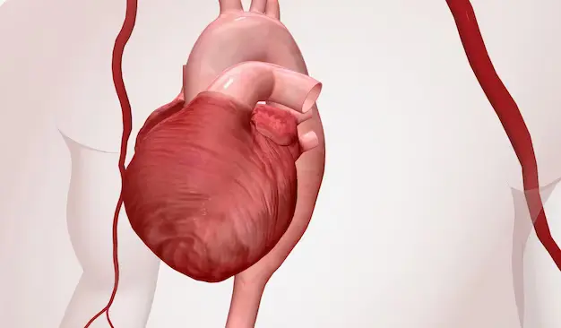
Physiological compensatory mechanisms lead to left ventricular hypertrophy and a transient increase in ejection fraction to tolerate the rise in ventricular blood volume. In the long term, the compensation will fail and lead to established heart failure, along with a marked reduction in ejection fraction and cardiac output.
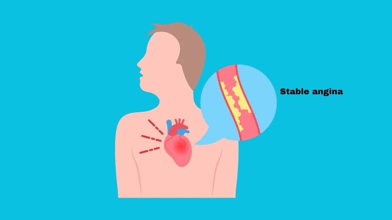
Furthermore, aortic regurgitation will affect blood perfusion through arteries, including coronary arteries, which contribute to myocardial ischemia. In addition, severe aortic regurgitation may participate in mitral valve regurgitation, which can predispose to cardiac arrhythmia.
Diagnosis and Physical and Clinical Findings
At Acute Aortic Regurgitation:
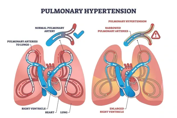
- Systemic hypotension and pulmonary hypertension are observed.
- In aortic dissection cases, pulse and pressure differences between arms and echocardiogram (ECG) ischemic changes are detected.
- Heart sound changes may be noticed: S1 will be soft or absent due to early closure of the mitral valve, S2 will be prolonged due to pulmonary congestion, and S3 will be heard.
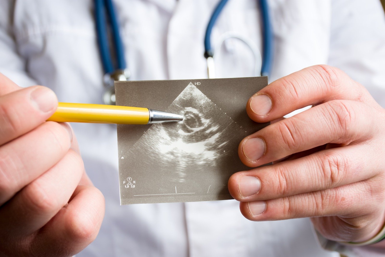
- Echocardiography (ECHO) can examine the aortic valve and show aortic root dilation.
- Trans-esophageal echocardiography (TEE) and computed tomography (CT) are effective for the diagnosis of aortic dissection and infectious endocarditis.
- Aortography or color Doppler can be used in stable patients for aortic dissection identification.
At Chronic Aortic Regurgitation:
- Pulse pressure is increased, so Traube's sign and Duroziez's murmur are noticed.
- Echocardiography would indicate left ventricular hypertrophy and ejection fraction reduction.
ACC / AHA Aortic Regurgitation Staging
It involves patients with a rheumatic history of infectious endocarditis or abnormal valves but without any evidence of regurgitation or clinical symptoms.
Structural valve abnormalities and mild to moderate regurgitation are identified but without any clinical symptoms.
Structural valve changes and severe regurgitation are detected. In addition to severe left ventricle dilation or alert systolic function, there are no clinical symptoms.
The patient is complaining of heart failure-related symptoms, along with structural valve abnormalities and severe regurgitation.
Management
-
Aortic valve repair or replacement
It is indicated as an emergency treatment for patients with severe acute aortic regurgitation. Also, in cases of paravalvular abscess due to infectious endocarditis
Mechanical valves have an extended efficacy duration but are associated with a higher thromboembolism risk when compared to bioprosthetic valves.
Intra-aortic balloon use is contraindicated since it may worsen the condition if inflation occurs during heart diastole.
-
Emergency surgery for ascending aortic grafting with or without aortic valve replacement
is indicated if both aortic regurgitation and aortic dissection are diagnosed.
Beta-blockers must be used with caution to avoid worsening hypotension due to reflex tachycardia inhibition.
-
Intravenous vasodilators (Nitoprusside) and Ionotropic (Dobutamine) use
They are used to stabilize the patient if the surgery is delayed. Vasodilators can reduce afterload while Dobutamine works on B1 to maintain cardiac output and on B2 for the vasodilation effect.
They are recommended for hemodynamically stable patients if aortic regurgitation is caused by infectious endocarditis. Also, antibiotics must be prescribed as a prophylaxis before dental procedures.
References


.webp)
.webp)
.webp)
.webp)
.webp)

.webp)