Premature Atrial Contractions (PACs)
Definition
Premature atrial contractions, also commonly referred to as atrial premature complexes, are ectopic heartbeats that originate from the atrium and could be linked to other atrial arrhythmias and associated with structural heart diseases.
Pathophysiology and etiology
They may be associated with the administration of caffeine, stimulants, adrenergic agonists, and smoking, induced by fever during acute infection, and linked with electrolyte disturbances, hyperthyroidism, hypertension, heart failure, acute myocardial infarction, cardiac amyloidosis, and sarcoidosis.
They may be generated from any atrial region. They emerged as a result of structural changes and dilation of the atria that led to reentry circuits and irregular impulse formation and conduction. They are usually benign, but they can trigger other tachyarrhythmias, such as atrial fibrillation and flutter, supraventricular tachycardia, and ventricular fibrillation.
Clinical presentation
Patients could be asymptomatic, but others could complain of palpitations and irregular heart rhythms and describe the feeling as a temporary halt or pause in their heartbeat followed by powerful heartbeats. Also, it may be induced following exercise.
Diagnosis
Secondary causes must be excluded, such as anemia, hypoglycemia, hypovolemia, stimulant intake, anxiety, panic attacks, and other structural cardiac or arrhythmic disorders.
Electrocardiography would show a narrow QRS followed by a pause when electrical signals are conducted through normal pathways. On the other hand, the P wave may be imposed over the T wave due to a pause when signals are stopped or blocked.
Premature Ventricular Contractions (PVCs)
Definition
Premature ventricular complexes are ectopic beats that are generated from ventricular sites below an atrioventricular node. They are usually benign but may be associated with reversible cardiomyopathies.
Pathophysiology and etiology
They could originate from ventricular tissues, such as right or left ventricular outflow tracts, atrioventricular valve annulus, left ventricular Purkinje fiber, adjacent structures, papillary muscles, or epicardium beside the aortic sinus of Valsalva, without coexisting structural heart diseases. In addition, they can emerge from infarcted tissue or the epicardium of dilated cardiomyopathy. Moreover, they may be idiopathic.
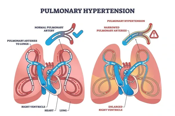
They could occur through several mechanisms: reentry circuits triggered beats, and enhanced automaticity. They can be linked with stimulant administration, pulmonary hypertension, obstructive sleep apnea, thyroid and adrenal gland disorders, myocarditis, cardiac amyloidosis, and sarcoidosis. In addition to congenital heart diseases and arrhythmias.
Clinical presentation
Some patients may be asymptomatic, while others may present with palpitations, shortness of breath, chest pain, fatigue, syncope, and pulsation in the head or neck. Also, dysphagia and dry cough may present in rare cases. Symptoms may be triggered by exercise, emotional stress, lying on the side, and food.
Diagnosis
Patient family and medical history, physical examination, and echocardiography are important for the diagnosis and exclusion of other medical conditions.
Electrocardiography with or without a Holter monitor and an exercise treadmill stress test are used to identify premature ventricular contractions that appear as a wide, weird QRS complex without a P wave, followed by an inverted T wave, and a longer than normal compensatory R-R interval. These beats could be monomorphic or polymorphic and appear as every other beat, every third beat, or every fourth.
Non-sustained ventricular tachycardia (NSVT)
Definition
(based on the 2017 AHA/ACC/HRS Guidelines for the Management of Patients with Ventricular Arrhythmias and the Prevention of Sudden Cardiac Death)
It is common but not clearly understood: arrhythmia. It is identified as an elevated heart rate >100 bpm with >=3 sequential ventricular beats that last for < 30 seconds. Usually, when it is detected in patients with structural heart diseases or during exercise, it can be used as a predictable marker for sustained ventricular tachycardia and sudden cardiac death.
Pathophysiology and etiology
It may have occurred to healthy people. However, if episodes are recurrent, then the existence of secondary causes must be evaluated, such as hypoxia, anemia, and electrolyte disturbances. Moreover, any accompanying structural heart diseases, such as heart failure and myocardial ischemia, must be identified.
Clinical Presentation
Some patients may present with palpitations, shortness of breath, and chest pain that can develop into syncope. Symptoms are usually dependent on the rate and duration of arrhythmia.
Diagnostic consideration
It is associated with widening of QRS > 120 ms and loss of coordination between atrium and ventricle contractions (known as atrioventricular dissociation), which presented with variable blood pressure and heart sounds, in addition to canon A wave with jugular vein pulsation.
References


.webp)
.webp)
.webp)
.webp)
.webp)
.webp)

.webp)