A pulmonary valve is a heart valve that consists of three thin leaflets. It is located anteriorly to the other valves, based on the superior view of the heart. It is a semilunar valve that connects between the right ventricle and the pulmonary artery, where blood flows through to the lungs to release cellular carbon dioxide waste and get saturated with inspiratory oxygen. Many congenital and acquired conditions may affect the structure or function of the pulmonary valve.

Pulmonary Valve Stenosis
It is the most common pulmonary valve disease and is associated with around 80% of right ventricular outflow obstruction cases; where blood outflow from the right ventricle becomes reduced or prevented. It usually emerges from congenital malformations rather than acquired disorders. In addition, it may acquired as a result of Rheumatic fever, Homograft dysfunction, and Carcinoid syndrome.
In valvular pulmonary stenosis, the pulmonary valve takes on a dome shape during heart systole, as the cusps are thin, the commissures are fused, and the center opening is narrowed, but the valvular movement is preserved. Furthermore, in subvalvular pulmonary stenosis, the pulmonary valve may be dysplastic, as valvular cusps become thickened with inadequate mobility and an atypical presentation of the hypoplastic annulus and proximal pulmonary artery.
Also, supravalvular pulmonary stenosis may have resulted from functional obstruction at distal branches of the pulmonary artery or obstruction in the right ventricular conus arteriosus (infundibulum) outflow tract.
Clinical Presentation
It is a congenital malformation that is benign in most neonatal ages and has a positive prognosis and a longer life expectancy. On the other side, if the stenosis is severe, it will be clinically manifested at an early age, which usually worsens over time. Patients may present with dyspnea, fatigue, exertional angina, or cardiac arrest. Also, it may be associated with right ventricular heart failure and arrhythmia. Moreover, severe pulmonary valve stenosis in infants is associated with a high mortality risk.
Diagnostic findings
- Upon physical examination, a left parasternal heave as a result of right ventricular hypertrophy may be noticed. Also, heart and pulmonary sound auscultation usually reveal a spilt second heart sound as pulmonary valve closing is delayed, a systolic murmur at the left sternal border, a noticed fourth heart sound, and tricuspid valve regurgitation.
- Echocardiography can detect any structural valvular defects and determine stenosis severity based on flow gradient and velocity across the pulmonary valve.

Pulmonary Valve Regurgitation
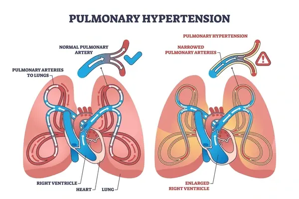
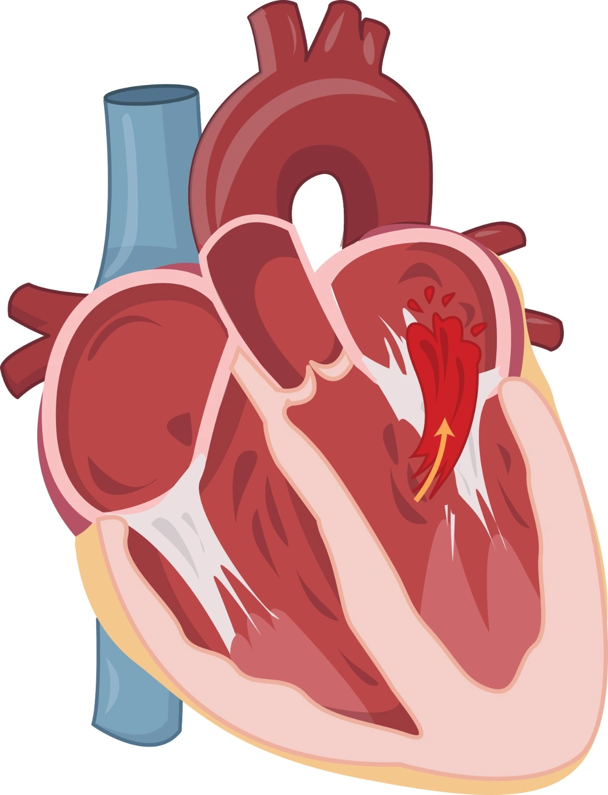
In this condition, the pulmonary valve is not appropriately closed, which leads to blood backflow to the right ventricle. Minimal Pulmonary Valve Regurgitation may be detected as a normal physiological condition. Usually, it is developed as a secondary complication of preexisting medical conditions, such as pulmonary hypertension or dilated cardiomyopathy, which affect cusps or dilate the ring of the pulmonary valve.
Etiology
- Pulmonary Hypertension is considered the most common cause associated with Pulmonary Valve Regurgitation.
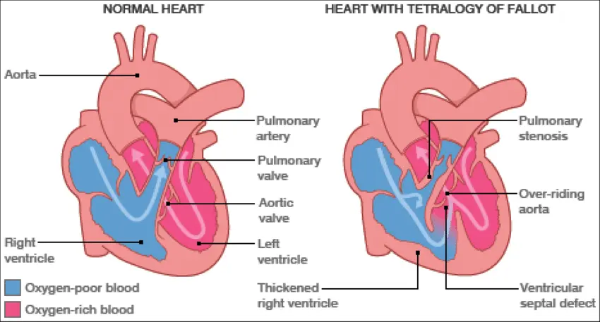
- A congenital heart defect such as Tetralogy of Fallot is considered a common cause.
- Infective Endocarditis and Rheumatic Heart Disease may contribute to severe Pulmonary Valve Regurgitation.
Clinical Presentation
Patients with Pulmonary valve Regurgitation are usually asymptomatic. Symptoms appear in later stages when the right ventricle is hypertrophic. Symptoms include fatigue, dyspnea, chest pain, and arrhythmias. Severe cases may be associated with palpable pulmonary veins, distended neck veins, peripheral edema, ascites, hepatosplenomegaly, light headache, and syncope.
Diagnostic considerations
- Heart sounds auscultation may detect third and fourth heart sounds at the left side of the mid to lower sternum border and heart murmurs at the second and third intercostal spaces on the left side during early heart diastole, which are more noticeable during inspiration.
- Jugular vein pressure is usually elevated.
- Echocardiography can identify Pulmonary Valve regurgitation and determine its severity. In addition, it may detect the underlying primary cause.

Pulmonary Valve Atresia
It is a birth defect where the pulmonary valve is not formed and a solid tissue takes its place, causing a blockage of blood outflow into the pulmonary circulation. Therefore, blood moves through alternative pathways such as the patent foramen ovale and ductus arteriosus to bypass the obstruction. It may be associated with ventricular septal defects and is considered a critical congenital valvular disease that needs urgent surgical intervention.
References


.webp)
.webp)
.webp)
.webp)
.webp)

.webp)