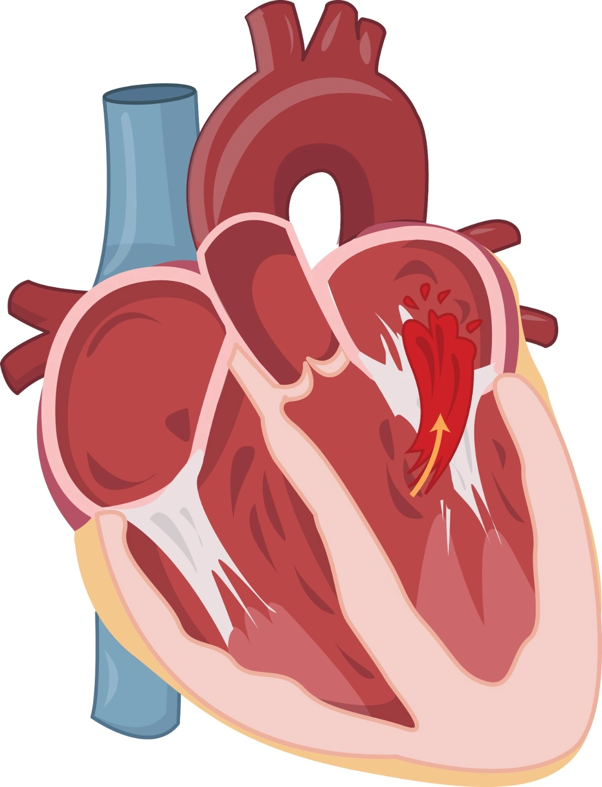
Aortic regurgitation is a common valvular heart disease where aging, uncontrolled Hypertension, and cardiac tissue infection may affect the aortic valve, leading to abnormal backward movement of oxygenated blood from the aorta into the left ventricle during ventricular diastole. As a result, the left ventricle wall will become hypertrophic as a compensatory mechanism to eject the whole blood from the left ventricle, but the compensatory mechanism may fail over time, leading to the development of Heart Failure.

- Blood pressure in the extremities
A positive Hill's sign when the systolic blood pressure difference between the upper and lower extremities exceeds 60 mmHg may be associated with aortic regurgitation.
- Inspection and Palpation of chest wall
It is done using fingers or palms while the patient is sitting up, leaning forward, or lying in the left lateral decubitus position, in which the heart is closer to the chest wall. Many findings associated with aortic regurgitation may be noted, including:
- Pulse pressure is usually elevated above 80 mmHg.
- Point of maximal intensity (PMI), -which is the point where the heart pulse is the strongest and is located at the apical point of the heart in the left intercostal space right below the nipple, may be found in more inferior and lateral positions in cases of chronic Aortic Regurgitation if the heart is dilated.
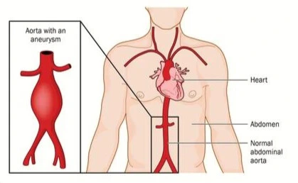
- A big pulsation at the sternum notch and right upper intercostal spaces may be noticed if Aortic Aneurysm is the underlying cause of Aortic Regurgitation.
- A corrigan-bounding pulse in the carotid artery due to the up and down movement of blood through the carotid artery may be noticed.
- Watson water hammers bounding a pulse in a radial, ulnar, or brachial artery may be noticed, especially when the patient's arm is lifted.
- Quincke’s capillary pulse on the patient's nail buds and lips may be noticed.
It is done when the patient is relaxed and sitting up, leaning forward during the end of the expiration. Also, the isometric handgrip cardiac maneuver can be used to increase cardiac afterload and make the Aortic Regurgitation murmur louder.
- Auscultation to the lower sternal border in the left third or fourth intercostal space may detect decrescendo-blowing murmurs during heart diastole. If it is noticed louder on the right side, it may indicate aortic dilation.
- Auscultation to the heart apex at the left fifth intercostal space using the bell of a stethoscope may detect Austin Flint murmur, which is a rumbling diastolic murmur associated with functional Mitral valve Stenosis that results from severe Aortic Regurgitation.
- Auscultation of the aortic region at the upper sternal border may detect a systolic outflow murmur, which may be associated with an Aortic Aneurysm that induces Aortic Regurgitation.
- S3 gallop may be detected when systolic heart failure develops due to chronic aortic regurgitation.
- Laboratory tests
- CBC with differential and culture tests are essential if infectious endocarditis is suspected.
- Serology tests may identify underlying Rheumatological disorders.
It can detect any hypertrophy in heart cells, volume overload associated with left ventricular dysfunction as a notable Q wave, and electrical conduction abnormalities.
During Acute Aortic Regurgitation, an increase in pulmonary venous flow pattern is noticed. In Chronic Aortic Regurgitation, cardiomegaly and aortic root protruding are noticed. It can also identify aortic valve calcification and pulmonary congestion.
-
- Transthoracic Echocardiogram
It must be performed on all patients with suspected Aortic Regurgitation. It is associated with high specificity and sensitivity results. It can be used to assess the following:
- Heart champers structural changes and function, including ejection fraction, for heart failure diagnosis.
- Aortic valve structure and motion to identify aortic regurgitation and determine its severity are based on left ventricle outflow tract central jet width, vena contracta width, regurgitant fraction and volume, and effective regurgitant orifice area.
- Vegetation exists, which is associated withinfectiouse Endocarditis.

- Angiography during cardiac catheterization
It can be performed to determine Aortic Regurgitation severity using a grading system.

References


.webp)
.webp)
.webp)
.webp)
.webp)
.webp)