Overview
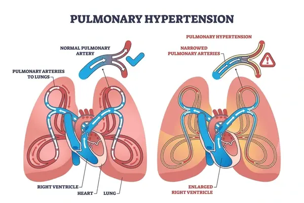
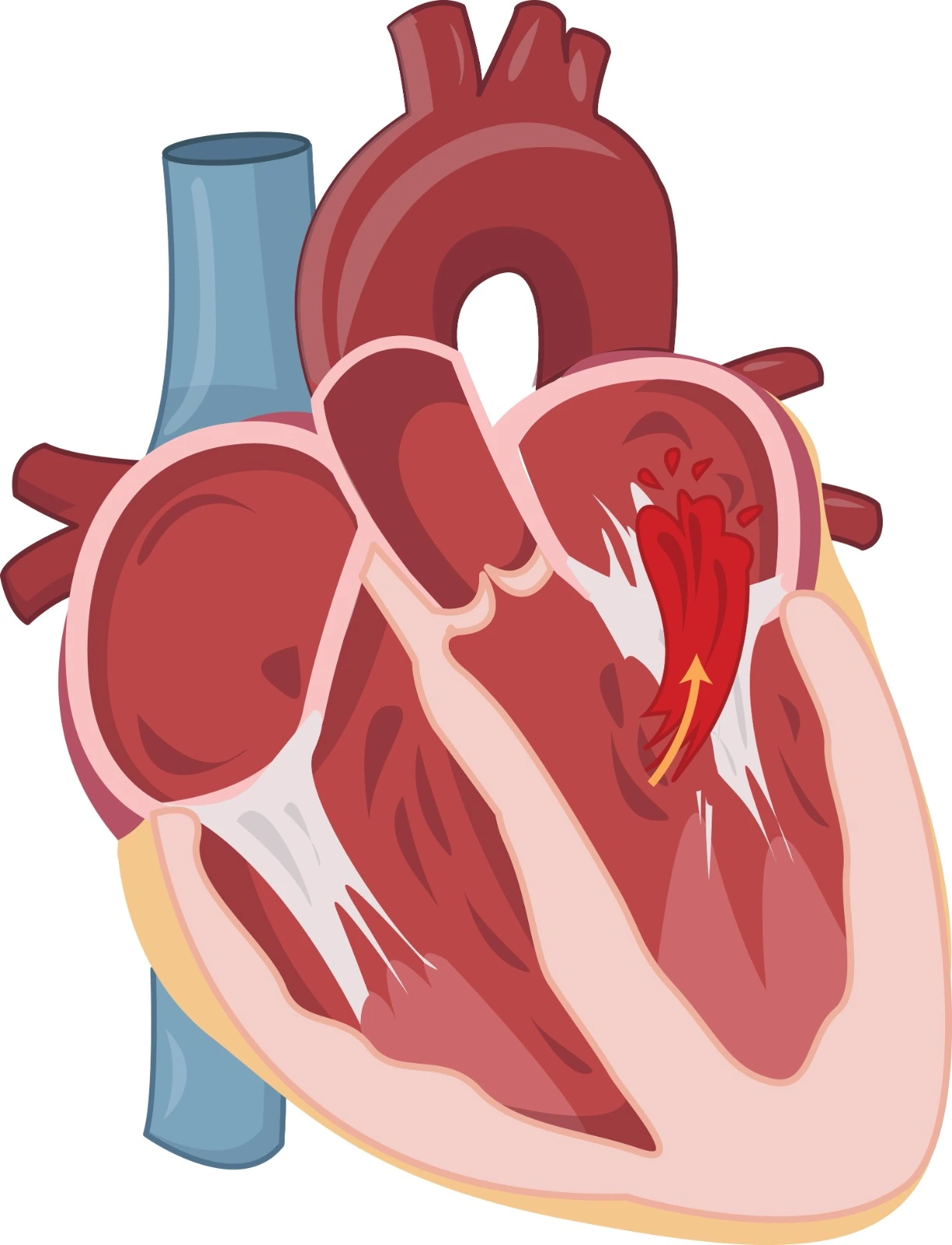
Mitral stenosis is the blockage of blood flow from the left atrium to the left ventricle. It is most commonly caused by rheumatic heart disease, but sometimes it can be non-rheumatic calcific mitral stenosis. Progression of the disease leads to disabling symptoms (e.g., dyspnea, hemoptysis, thromboembolism, pulmonary hypertension, and right-sided heart failure).
Etiology:
Rheumatic heart disease is the most common cause of valve disease worldwide. Its prevalence is declining in high-income countries and is higher in low and middle-income countries. Rheumatic MS is more common in females than in males.
The presentation of the disease differs from one region to another; patients from high-prevalence regions usually present at a young age with no calcification but with commissural fusion, whereas patients from low-prevalence countries present at old age with calcification, commissural fusion, sub-valvular involvement, and usually have another cardiac disease, eg: atherosclerotic cardiovascular disease.
Pathophysiology
As MS has many etiologies, the pathophysiology differs from one to another. It also differs based on the factors that contribute to the progression and the hemodynamic and structural consequences.
The thickening and immobility of the mitral valve leaflets lead to a blockage of the blood flow from the left atrium to the left ventricle, leading to increased pressure in the right atrium.
Rheumatic Mitral Stenosis:

Clinical Manifestations
Note that these clinical manifestations might be exacerbated by certain precipitants, such as exercise or stress.
- Dyspnea, the most common manifestation, occurs due to elevated left atrial pressure and pulmonary venous hypertension.
- Fatigue: This happens due to decreased cardiac output and the development of heart failure.
- Hemoptysis: due to an elevation in pulmonary pressure and vascular congestion.
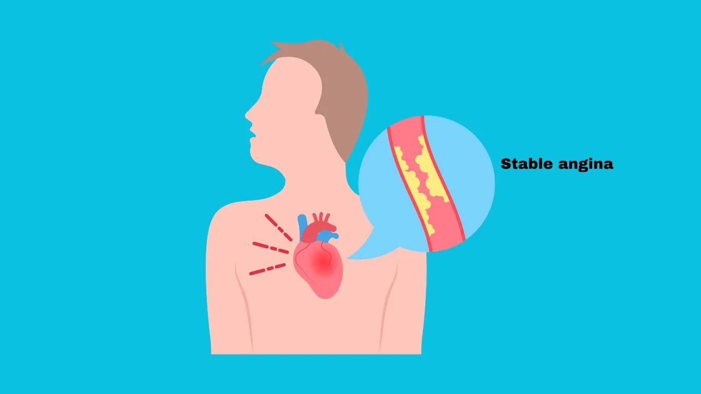
- Chest pain is a rare clinical manifestation.
Physical examination
- Mitral facies
- Crackles due to pulmonary edema
- Signs and symptoms of right-sided heart failure. (eg: ascites)

Diagnosis
Echocardiography is considered the ultimate imaging modality in patients with rheumatic mitral stenosis. To establish a diagnosis, a transthoracic echocardiogram (TTE) is indicated for patients with signs and symptoms and to assess their need for percutaneous mitral balloon commissurotomy (PMBC). If PMBC is considered, TTE should be done to exclude the presence of a left atrial thrombus.
Exercise: The hemodynamic threshold of exercise testing is helpful in symptomatic patients with exertion (dyspnea) and a resting mitral valve area > 1.5 cm2. It helps identify patients who would benefit from PMBC. These patients usually have an increase in pulmonary artery wedge pressure (>25 mmHg) during exercise testing or cardiac catheterization or a mean mitral valve gradient >15 mmHg.
Cardiac catheterization can be applied when non-invasive methods are inconclusive, or it can be used alongside echocardiography to monitor hemodynamics during a PMBC procedure.
Mitral Stenosis Staging

Rheumatic MS Medical Therapy:
- Rheumatic MS and AFIB, a previous embolic event, or left atrial thrombus: Anticoagulant and vitamin K antagonist (e.g., Warfarin).
- Rheumatic MS and AFIB with a rapid ventricular response: rate control
- Rheumatic MS with normal sinus rhythm WITH resting or exertional tachycardia: rate control

Non-rheumatic calcific mitral stenosis:
Although it is rare, it can be detected in older adults. It happens due to calcification of the mitral annuals that extends to leaflet bases; therefore, the annuals will become narrowed and will lead to rigid leaflets with no commissural fusion.
Because these patients are usually older adults with comorbidities, the indication of any intervention for any patient with calcific MS differs from those with rheumatic MS, and the patient should be highly symptomatic. Symptoms can be managed using dieresis and rate control unless they are very severe and disabling.
Limitations of intervention:
- There is no role for PMBC or surgical commissurotomy.
- Calcification is very challenging for the surgeon to place a prosthetic valve.
- Patients are usually at high surgical risk due to old age and comorbidities.
References:
- https://www.uptodate.com/contents/rheumatic-mitral-stenosis-clinical-manifestations-and-diagnosis
- https://www.jacc.org/doi/pdf/10.1016/j.jacc.2020.11.018?_ga=2.101304452.482784417.1698062847-100308460.1697546937
- https://www.uptodate.com/contents/pathophysiology-and-natural-history-of-mitral-stenosis
- https://www.ncbi.nlm.nih.gov/pmc/articles/PMC10504831/
- https://www.uptodate.com/contents/rheumatic-mitral-stenosis-overview-of-management


.webp)
.webp)
.webp)
.webp)
.webp)
.webp)