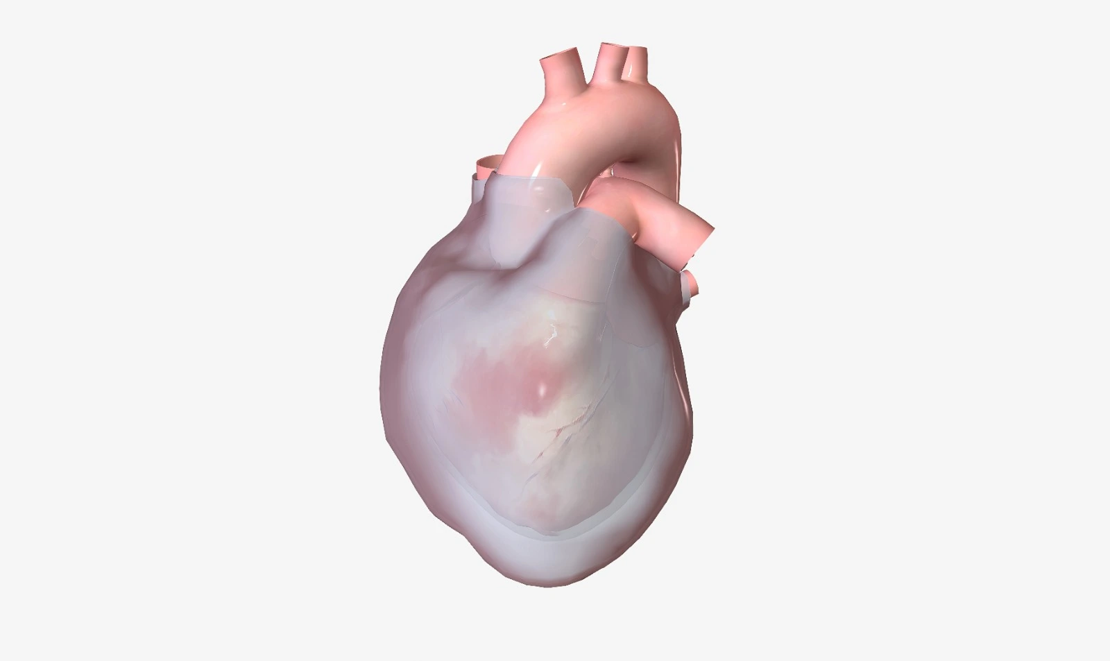
The pericardium is an outer envelope that covers the heart. The pericardial sac normally contains 15 to 50 ml of fluid. Pericardial fluid may excessively accumulate inside the pericardial sac and develop pericardial effusion. If intrapericardial pressure is elevated rapidly or is associated with massive pericardial volume expansion, it will lead to symptomatic, life-threatening cardiac tamponade.
Pathophysiology
Accumulated pericardial fluid could be transudate fluids, as in the case of obstruction in lymphatic drainage, or exudate fluids originating from viral infections such as HIV, bacterial infections such as Tuberculosis and fungal or parasitic infections in rare conditions, inflammatory and autoimmune disorders, and malignancy from lung, breast, leukemia, and lymphoma, or blood from acute aortic dissection. In addition to uremia in renal failure, dyslipidemia, myocardial infarction-induced free wall rupture, chest wall trauma, and medications
Cardiac tamponade is developed due to malignancies, which are considered the main leading cause of acute pericarditis and conditions associated with severe pericardial effusion and elevated intrapericardial pressure. Also, it's more common in males than females. It is a medical emergency syndrome that occurs when accumulated fluid in the pericardial space elevates intrapericardial pressure and impedes ventricle relaxation. Therefore, higher filling pressure is required to overcome pericardial pressure. After a while, ventricular filling will drop and result in reduced cardiac output, which could lead to hemodynamic instability and shock and be associated with a higher mortality risk.
Clinical presentation
Pericardial effusion could be rapidly developed, in which even a small abnormal amount of fluid could contribute to symptoms. Otherwise, it could be slowly developed, where a larger fluid amount—reaching 2 liters—may be accumulated before developing symptoms.
Pericardial effusion symptoms are usually dependent on underlying causes and associated complications, which include:
- Chest pain that becomes worse in the supine position.
- Cardiac palpitations
- Cough and shortness of breath.
- Hiccups and hoarseness
- Anxiety, a light headache, and syncope
Cardiac tamponade clinical presentation is based on the underlying cause and includes:
- Weight loss, fatigue
- Fever
- Trauma
- Tachycardia and palpitations
- Tachypnea and dyspnea
- Hepatomegaly
- Dizziness
- Restless body movements
Physical examination
Upon physical examination, some findings may be associated with pericardial effusion and cardiac tamponade, such as:
- Becks triad is identified in acute pericardial effusion and reflects acute compression.
Where blood pressure is decreased, jugular vein pressure is distended, and heart sounds are muffled,
- Pulsus paradoxus is detected with Cardiac Tamponade as systolic pressure falls more than 10 mmHg during inspiration.
During inspiration, intrathoracic pressure is decreased due to volume expansion, which induces right ventricle filling and subsequent septal bulging and shifting into the left, so left ventricle filling and cardiac output are reduced— as an obstructive shock —and the left ventricle cannot expand as compensation due to the pressure of pericardial effusion.
- Attenuated or revoked Y descent in the atrial waveform
Jugular vein pressure is elevated as ventricular filling during heart diastole is reduced and intrathoracic pressure is increased.
It’s a dullness to percussion in the left lower lung due to lung compression by a large pericardial effusion.
Diagnosis
Diagnosis is established based on patient symptoms and affirmed with echocardiogram findings:
It must be done for all suspected patients, as it’s the gold standard for pericardial effusion diagnosis and quantification.
It's used for cardiac tamponade diagnosis and specification of location, size, and hemodynamic effect of effusion.
The degree of effusion is based on the space between pericardial layers during diastole.
- A small pericardial effusion is < 10 mm.
- Moderate pericardial effusion is 10–20 mm.
- A large pericardial effusion is ≥ 20 mm.
- The chest X-ray is not a specific or sensitive imaging test, but it can be used to detect pericardial effusion as the heart will appear in a water bottle shape.
- Computed tomography (CT) and magnetic resonance imaging (MRI) may be able to identify pericardial effusion.
- Blood tests must be ordered to evaluate markers of inflammation and myocardial injury.
References


.webp)
.webp)
.webp)
.webp)
.webp)
.webp)

.webp)