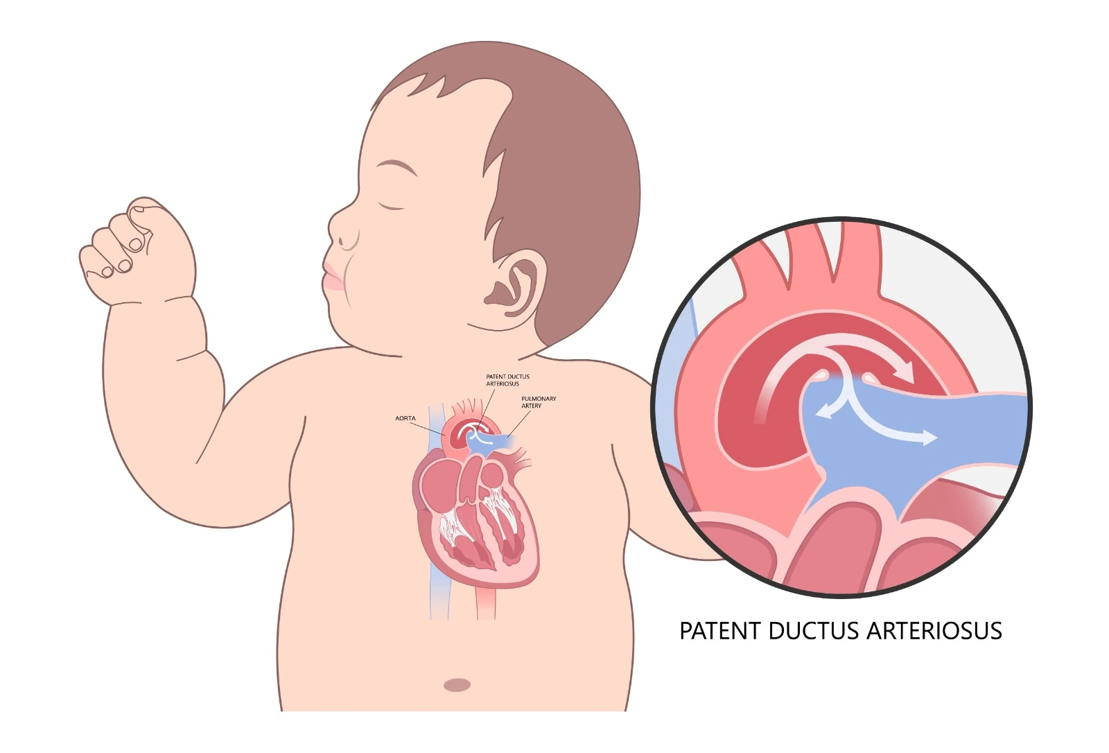
Fetal heart and blood vessel formation occurred during the first trimester of pregnancy. Ductus arteriosus is a normal small blood vessel that connects the aorta and pulmonary artery. It is normally formed during fetal life and contributes to around 60% of cardiac output. When oxygenated blood from the maternal placenta reaches the right atrium and ventricle and then is pumped into the pulmonary artery, it will bypass pulmonary circulation by allowing the oxygenated blood to directly move through the ductus arteriosus into the aorta and then to the systemic circulation.
The ductus arteriosus is maintained open during fetal life under the effect of prostaglandin E2, then it is normally closed shortly after birth when the ductus arteriosus wall is contracted as oxygen is inhaled and pulmonary vascular resistance is elevated, which allows blood movement through pulmonary circulation when normal air breathing is initiated, and it will be known as the ligamentum arteriosum (arterial ligament).
Pathophysiology
In the case of Patent Ductus Arteriosus, the connection between the proximal descending part of the aorta and the left pulmonary artery remains open after birth, which forms a left-to-right shunt. So, the blood will flow from the aorta to the pulmonary artery and then into the pulmonary circulation instead of moving to the systemic circulation. As a result, blood movement through the patent ductus arteriosus will elevate the blood pressure in the pulmonary circulation.

Etiology
The exact underlying causes of patent ductus arteriosus are unknown, but many risk factors may be associated with the condition:
- Patent ductus arteriosus is a common condition in premature infants, while it’s a rare condition in full-term babies, where it's usually associated with other congenital heart defects.
- A family history of congenital heart defects may be associated with patent ductus arteriosus.
- Dawn syndrome patients are at higher risk of patent ductus arteriosus.
- Getting infected with Rubella or German Measles during the first trimester of pregnancy increases the risk of fetus congenital heart defects.
- Being born at high altitudes with low atmospheric oxygen levels may be associated with a higher risk of patent ductus arteriosus.
- Female gender and low birth weight may be risk factors.
Clinical Presentation
In mild cases of patent ductus arteriosus, patients may not experience any symptoms. In severe cases, affected children may present with shortness of breath, tachypnea, tachycardia, fatigue, poor feeding, which affects their growth, and pneumonia. Persistent, untreated severe patent ductus arteriosus may lead to pulmonary hypertension and Eisenmenger syndrome, where permanent lung damage occurs. Moreover, the heart may be affected when heart failure and infectious endocarditis are diagnosed.
Diagnosis
Although Patent Ductus Arteriosus is usually diagnosed during childhood, it may be delayed until adulthood when it is mild. A clinical diagnosis can be done through:
- Physical examination and heart auscultation can detect heart murmurs.
- Routine blood tests to evaluate general health. Also, polycythemia may be noticed with other congenital heart defects.
- Arterial blood gases can reveal acidosis that is connected to pulmonary hypertension, while hypercapnia and hypoxia are noticed if patent ductus arteriosus induces heart failure.
- Doppler echocardiography can identify a high-velocity turbulent flow in the pulmonary artery that is diagnostic of patent ductus arteriosus. Also, left atrium and ventricle enlargement are detected in severe cases.
- Chest radiography can detect pulmonary vessels and left heart chamber enlargement, along with pulmonary congestion, which may develop. In addition, the ductus arteriosus may be calcified in advanced-age patients.
- Cardiac angiography may be used to observe the shunt and pulmonary artery pressure and for other cardiac disease diagnoses.
A: Echocardiogram; (DAO) Descending Aorta, (MPA) Main Pulmonary Artery / B: Doppler image

Classification of Patent Ductus Arteriosus
The Krichenko classification based on angiography:
- Type A (conical), aortic ampulla, and constriction near the pulmonary artery is defined.
- Type B (window), huge but very short ductus arteriosus
- Type C (tubular), no identified constriction
- Type D (complex), multiple constrictions are detected.
- Type E (elongated), constriction is remote from the tracheal anterior edge.

References

.webp)
.webp)


.webp)


.webp)

.webp)


.webp)