A physical examination is an essential part of the clinical diagnosis process. Stethoscopes are among the tools that can be used during the examination. They are used to assess internal body sounds from the heart, lungs, and abdomen by using one or both of two chest pieces: a small concave bell that allows listening for low-frequency sounds and a large flat diaphragm that allows listening for high-frequency sounds. Auscultation of heart sounds has a big role in blood pressure measurement, heart rate and rhythm assessment, and valvular disorder detection.
The normal heart sound represents a complete heart cycle, which consists of S1 and S2. The duration between S1 and S2 represents cardiac systole. While the gap between S2 and S1 represents cardiac diastole.
First heart sound (S1)
It is a normal heart sound, known as "Lub", resulting from blood recoil against closed atrioventricular valves at the end of diastole, just before systole and the carotid upstroke pulse. Closure of the mitral valve (M1) can be loudly heard on top of the heart apex, immediately followed by tricuspid valve closure (T1), which is best heard at the 4th intercostal space over the left side of the sternum.
It is considered a high-frequency sound, and acuity is dependent on atrial emptying rate, ventricular pressure elevation rate, trans-valvular rate and gradient, and valvular competency and ability to widely open and quickly complete closure, which are reflected in the PR interval.
Loader S1 may be associated with:
- Higher trans-valvular gradient due to tricuspid or mitral valve stenosis.
- Elevated trans-valvular flow as in patent ductus arteriosus (left to right shunt)
- Tachycardia induces rapid ventricular contraction due to hyperthyroidism, anemia, hypoglycemia, medications, and exercise.
- Short PR interval by pre-excitation; Wolf Parkinson White Syndrome (WPWS)
Lower S1 may be associated with:
- Severe mitral valve stenosis is associated with valvular immobility.
- Tricuspid or mitral valve regurgitation occurs when fibrosis leads to inappropriate valve closure or if the trans-valvular gradient is disturbed.
- Ventricular dysfunction is induced by cardiomyopathy, myocardial infarction, and medications such as beta blockers.
- Left bundle branch block that delays left ventricle contraction.
- Prolonged PR interval due to heart block.
- Wide distance through the chest wall, as in the case of pericardial or plural effusion and obesity.
Split S1 appears when tricuspid valve closure is delayed or mitral valve closure happened earlier. It may be associated with:
- Right bundle branch block.
- Ebstein anomaly, which affects the tricuspid valve
- Premature left ventricle contraction
- Atrial septal defect
Variable S1 may be due to atrial tachycardia, arrhythmia, or a complete heart block.
Second heart sound (S2)
It is a normal heart sound with high frequency, known as "Dub", related to blood movement against closed semilunar valves; as aortic valve closure (A1) that can be heard above the second intercostal space at the right side of the sternum, just before pulmonary valve closure (P1) briefly after the end of cardiac systole.
The intensity is affected by aortic and pulmonary valve status and is directly related to aortic and pulmonary artery pressure.
Wide physiological splitting S2 is produced due to the hang-out period that represents the interval between pressure crossover and actual valve closure, which is shorter in the aorta compared to the pulmonary artery. So, it is more noticeable during inspiration due to right ventricle volume augmentation.
Splitting S2 during expiration is related to a delay in pulmonary valve closure, as in the case of conduction delay in the right ventricle, pulmonary artery, or valve stenosis. Also, it may occur due to early aortic valve closure, as in mitral regurgitation or ventricular septal defect.
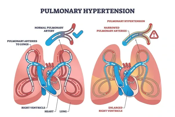
Splitting S2 could be fixed when respiration doesn’t have an impact on right ventricle volume, as in atrial septal defect, right ventricular heart failure, and pulmonary hypertension.
Reversed splitting S2 occurs when the aortic valve is closed after the pulmonary valve due to a conduction delay in the left ventricle or obstruction in the left ventricle outflow.
Single S2 is rare but may be associated with pulmonary or aortic valve severe stenosis and immobility.
Third heart sound (S3)
It is a low-frequency heart sound, best heard at the heart apex while the patient is in the left lateral decubitus position with breath held at end-expiration or at the left lower sternal border while in the supine position. It can be detected at early diastole, just after S2, due to the rapid movement of large blood volumes from the atrium to the ventricle, which causes vibrations along the ventricular walls. Ventricular compliance may be normal or reduced due to ventricular dysfunction; then it will be affected by even a normal blood volume movement, and in this case, it is known as ventricular gallop.
- It is considered physiological in children and young adults.
- It is pathological in patients over the age of forty and is associated with:
- Diastolic or systolic ventricular dysfunction
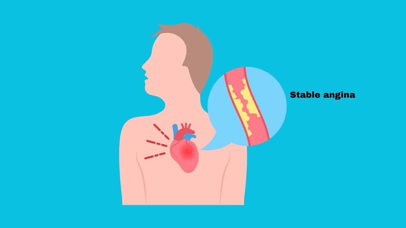
- Coronary artery disease
- Hypertension.
- Valvular Regurgitation.
- Hyperthyroidism.
- Anemia
- Fever.
Fourth heart sound (S4)
It is a low-frequency heart sound, best heard at the heart apex while the patient is in the left lateral decubitus position at the end of expiration and at the left lower sternum border. It is audible at late diastole, just before S1.
- It may be considered normal in the elderly due to low ventricular compliance.
- It's pathological at any age if it is palpable, known as atrial gallop, and may be caused by:
- Enforced atrium contraction into the non-compliant ventricle.
- Elevated preload or afterload along with ventricular dysfunction.
- Pulmonary Hypertension.
- Pulmonary or aortic valve stenosis
- During an acute myocardial infarction
References


.webp)
.webp)
.webp)
.webp)
.webp)
.webp)