Blog | Aortic Stenosis (AS) Diagnosis | Cardiologymaster
...

Aortic Stenosis (AS) Diagnosis

...
Aortic Stenosis (AS) Diagnosis

Aortic Stenosis (AS) Diagnosis

Admin
Nov 19, 2023

Overview

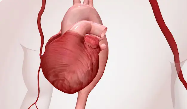
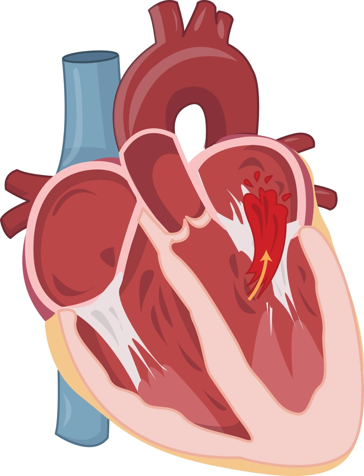
Aortic stenosis is a valvular heart disease that happens when the aorta is narrowed and cannot function fully, leading to a reduction in blood flow to the body.

Diagnosis

Symptoms:

  • Dyspnea with exertion is the most common symptom of AS. This happens due to diastolic dysfunction with LV filling pressure elevation during exercise and the inability of LV to increase cardiac output.
  • Dizziness and syncope are due to decreased cerebral perfusion.
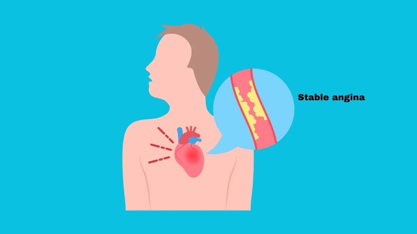
  • Angina pectoris with effort in severe AS, especially in patients with underlying coronary artery disease (CAD),

Physical Examination:

  • Carotid Pulse: "Parvus and Tardus" mean reduced amplitude and delayed occurrence of pulse, although the amplitude might be preserved in older adults. The delay can be detected by examining the apex and carotid artery at the same time. "Shuddering" or "Thrill" of the carotid artery might also be detected.
  • Precordial Palpation: Some patients have palpable S4 sounds, and a systolic thrill might be felt at the right intercostal space or the sternal notch, especially during full expiration while the patient is leaning forward.
  • Cardiac Auscultation:
  1. S2 is soft and single and may become paradoxically split in severe AS cases with LV dysfunction. Aortic closure (A2) is delayed and occurs at the same time as pulmonic closure (P2). In extremely severe cases, the aortic closure sound may disappear. A normal S2 split is a reliable finding to exclude severe AS.
  2. S1 is usually normal. When leaflets are stiff but compliant and mobile during the early stages of AS, an aortic ejection click might be heard after S1, especially with a congenital bicuspid valve.
  3. S4 might be produced due to vigorous left atrial contraction against a noncompliant, stiff ventricle.
  4. Systolic "ejection" murmur is best heard at the right intercostal space and transferred to the carotid artery at the same intensity, and any decreased intensity in the carotid artery might be a stenosis indication in that artery. A 4 or greater murmur might indicate severe stenosis. The murmur might be soft and undetectable in patients with low flow low gradient AS. In mild cases, the murmur is an early-peaking murmur, whereas in severe cases, it is late-peaking, This means that the timing of the murmur might detect the severity of the stenosis.

Echocardiography: is the essential key to diagnosis. It should be done when the BP is stable, and it depends upon the measurement of the mean pressure gradient, peak transvalvular velocity (Vmax), and valve area.

Categories

Category 

Mean gradient (mmHg)

Peak velocity (m/s)

Valve area (m2)

LVEF (%)

Stroke volume index (mL/m2)

Severity of Diagnosis

High-gradient AS

>=40

>=4.0

<=1 or <=0.6 cm2 /m2

NA

NA

Severe AS regardless of LV function and flow conditions

Low-flow, low-gradient AS with rEF

<40

NA

<=1

<50

<=35

Severe AS or pseudo-severe AS Can be detected using low-dose stress Dobutamine echocardiography:

pseudo-severe AS: increase in valve area to >1 and increased flow.

Low-flow, low-gradient AS with pEF

<40

NA

<=1

>=50

<=35

- Might be found in hypertensive, elderly patients with small LV sizes and marked hypertrophy, and in conditions associated with low stroke volume.


- Diagnosis of severe AS is difficult and requires accurate exclusion of other finding errors, the presence or absence of typical symptoms, LV hypertrophy, and normotensive or reduced LV longitudinal strain with no other cause.


- Additional important information can be obtained using CCT to detect calcification degrees (Agatston units):

  • Severe AS Highly likely:

Women > 1600, Men > 3000

  • Severe AS Likely: Women > 1200, Men > 2000
  • Severe AS Unlikely:

Women < 800, Men < 1600

Normal-flow, low-gradient AS with pEF

<40

NA

<=1

>=50

>35

Moderate AS


Additional tests:

  • Doppler velocity index (DVI): This may support evaluation when all other parameters are equivocal. A value of < 0.25 suggests that severe AS is highly likely.
  • Identifying patients with severe AS who are at risk of deterioration or premature mortality can be done through the assessment of global longitudinal strains when the threshold is 15%. 
  • Natriuretic peptides: can be used to determine other symptom sources in patients with multiple diseases that might contribute to these symptoms. It is also helpful in detecting high-risk asymptomatic patients who might benefit from early therapeutic interventions.
  • Exercise testing might be helpful to unmask asymptomatic patients with severe aortic stenosis and is also of benefit in detecting mean pressure gradients and alterations in LV function.
  • Diphosphonate and/or cardiovascular magnetic resonance are used when cardiac amyloidosis is suspected.
  • Coronary angiography is important before transcatheter aortic valve implantation (TAVI) and surgical aortic valve replacement (SAVR) to assess the need for revascularization.   

References:

  1. https://www.mayoclinic.org/diseases-conditions/aortic-stenosis/symptoms-causes/syc-20353139
  2. https://www.uptodate.com/contents/clinical-manifestations-and-diagnosis-of-aortic-stenosis-in-adults
  3. https://www.escardio.org/Guidelines/Clinical-Practice-Guidelines/Valvular-Heart-Disease-Guidelines
Share

Latest News