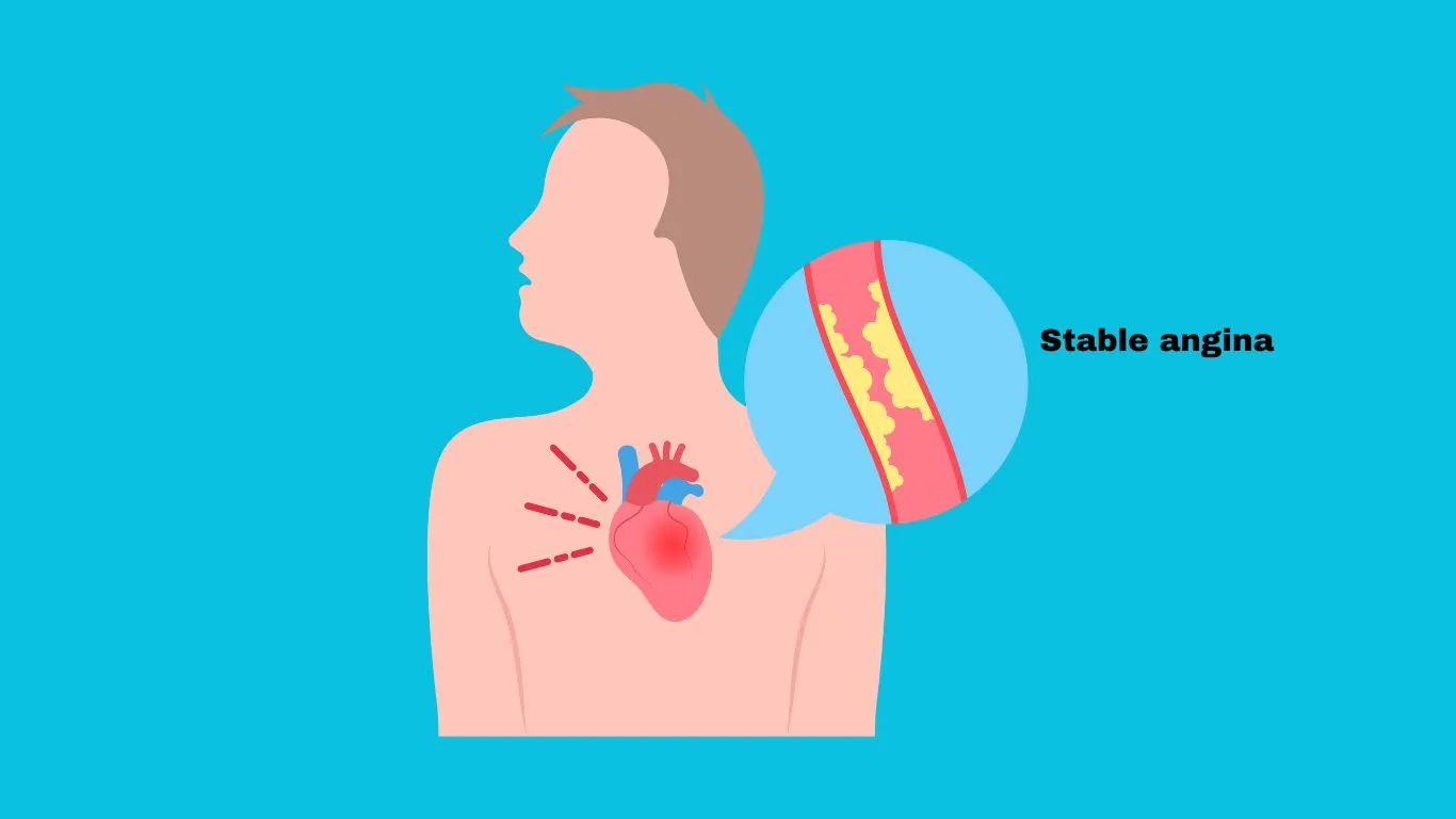
Stable Angina , which is also known as "Typical Angina" or "Angina Pectoris" is a manifestation of coronary heart disease that is responsible for major annual deaths. It is developed as a result of heart muscle ischemia due to a coronary artery narrowing that may be exacerbated to be significant enough to induce symptoms.
Pathophysiology
The coronary arteries are responsible for myocardial blood supply. Myocardial oxygen supply depends on coronary artery diameter and tone, heart rate, perfusion pressure and collateral blood flow, as myocardial oxygen extraction is limited due to low venous oxygen saturation. While myocardial oxygen demand is determined based on myocardial ionotropic and chronotropic functions, myocardial wall tension, and systolic blood pressure. Under normal conditions, coronary blood flow can be increased by several folds with maximal coronary dilation, which is known as coronary flow reserve (CFR).
Myocardial ischemia results from coronary blood flow hinder, which is induced by significant coronary artery stenosis that exceeds 50%, elevated coronary artery resistance, or reduced blood oxygenation.
Atherosclerosis, infections, and stress may cause disturbances in coronary arteries endothelial lining, which will affect its role in regulating vascular tone and preventing thrombus formation. As a result, the immunological response, coagulation cascade, and fibrosis will be activated, which will lead to coronary artery stenosis. Stenosis influence can be exacerbated by several factors which will increase myocardial oxygen demand that can't be matched by myocardial supply, which results in myocardial ischemia.
Subsequently, myocardial ischemia will activate chemosensitive and mechanoreceptive receptors, which will stimulate sympathetic impulses toward the cervical and thoracic spine, and each has a corresponding dermatome that is responsible for associated symptoms. Moreover, anginal symptoms are also linked with adenosine release and cardiac a1 receptors activation. Furthermore, myocardial-related metabolism process will be switched toward anaerobic metabolism which has a negative impact on mechanical and electrical myocardial function.
Clinical presentation
Patients with stable angina usually complain of diffused squeezing and pressure-like retrosternal chest pain, which may radiate to the arm, neck, jaw, shoulder, and back. It is usually triggered by physical activities, emotional stress, exposure to extreme weather, heavy meals, and smoking, and exacerbated by exertion. Symptoms usually last for a short period of time (<5 minutes), and are alleviated by rest or Nitroglycerine administration.
Diagnosis
Patients with anginal symptoms must seek medical advice to confirm stable angina diagnosis, determine the underlying cause, and exclude other medical conditions. In addition, angina pectoris diagnosis must not be delayed, as myocardial ischemia may be progressed into acute coronary syndrome with a high mortality risk.
- Physical examination is usually normal, but some findings may give a clue about the condition and the underlying cause.
- The Levine sign, where the patient holds a clenched grip over the sternum to describe chest pain, is a universal sign of ischemic chest pain.
- Xanthoma is a sign of lipid metabolism abnormality.
- Lack of peripheral pulse may indicate Atherosclerosis.
- Heart auscultation may reveal a fourth heart sound (S4) that is linked with left ventricular dysfunction or mitral regurgitation.
- Chest Radiography is typically normal in patients with stable angina, but it may reveal some accompanying complications.
- Exercise stress testing is the most commonly used diagnostic and prognostic test for stable angina.
- Stress Echocardiography is useful to evaluate localized ischemia, left ventricular function, cardiac dimensions, and heart valves.
- Computed Tomography is used for fast coronary artery calcium (CAC) scoring as a screening tool in asymptomatic patients, a predictive tool in symptomatic patients, and a prognostic tool in patients with confirmed stable angina.
- Computed Tomography Coronary Angiography (CTCA) is used to assess coronary artery stenosis.
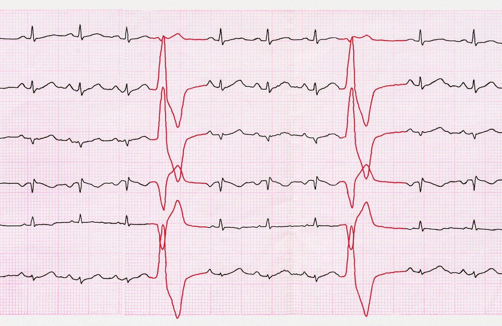
- Electrocardiography (ECG) can detect a >=1mm ST segment depression, 80ms away from the J point, during the angina attack in half of the patients with normal resting ECG. As resting ECG turns up normal in half of the patients with stable angina. So, exercise ECG is indicated after holding Beta Blockers for 48hrs, which reveal a >2mm ST depression that persists for 5 minutes after the end of exercise in case of ischemic heart disease that is associated with a poor prognosis.
- Invasive Coronary Angiography is the definitive test for the diagnosis and evaluation of coronary artery disease.
References
https://www.heart.org/en/health-topics/heart-attack/angina-chest-pain/angina-pectoris-stable-angina
https://www.ncbi.nlm.nih.gov/books/NBK559016/
https://emedicine.medscape.com/article/150215-overview#a1


.webp)
.webp)
.webp)
.webp)
.webp)

.webp)
